ZEN core is the universal software designed to operate any ZEISS light or electron microscope. Control your microscope with a single, standardized graphical user interface. This highly flexible software suite lets you perform exactly the microscopy workflows you need.
ZEN stands for ZEISS Efficient Navigation. ZEN core lets you obtain more meaningful information. It increases productivity, especially in multi-user labs and not only ensures data repeatability and reliability but also connects data from different modalities and locations. ZEN core is your ecosystem for seamless, end-to-end microscopy workflows.
Highlights
Image
Image Effortlessly and Configure the UI to Your Needs
Obtain images rapidly, control your instrument easily, and image all samples effectively. The GUI is common to any ZEISS light, scanning electron microscope, and focused ion beam SEM, a concept unique to ZEN core. And, once familiar with the standardized interface, you can easily switch between modalities without additional training effort. Users of all skill levels, especially in multi-user environments, will greatly benefit from the flexibility of this harmonized software. Enjoy enhanced productivity in your lab or imaging facility.
Control any ZEISS microscope and your ZEISS microscope camera.
Benefit from a user interface adaptable to the needs of your lab environment.
Adapt the configuration to your tasks.
Analyze
From Large Area Imaging to AI-based Analysis
Microscopists can rapidly execute advanced imaging tasks and perform post-processing analysis with ZEN core. When working with light microscopes, you will benefit from the consistency of advanced and repeatable workflows by using built-in automated image acquisition routines. For ZEISS SEMs and FIB-SEMs, any user can run standard workflows.
Obtain images of large areas using the functionalities for automated panorama acquisition or tiled image acquisition.
Easily execute automated image segmentation based on machine learning or deep learning algorithms, such as phase analysis, layer thickness measurement or grain boundary analysis.
Address typical research and quality control questions using application-specific toolkits e.g., multi-channel acquisition, GxP or technical cleanliness analysis.
Connect
Smart Solutions for Connected Microscopy
Keep your valuable analysis data together across instruments, laboratories, and locations. ZEN core provides smart solutions for connected laboratory environments. The single GUI of ZEN core bridges different applications from all kinds of microscopy modalities and improves multi-modal data integrity.
Toolkits for correlative microscopy, data management and database connectivity features help you to:
Connect all imaging and analytical data between multiple locations and instruments.
Manage users with different skill levels in multi-user environments.
Explore and visualize your data, organize them across scales and imaging modalities via mobile access.
Finally, ZEN core´s powerful documentation functionality creates reports in a fully automated manner.
ZEN core is an innovative software platform with a clear and intuitive operating concept and sophisticated functions for correlative microscopy, suitable for all types of imaging systems as it can process data from different microscope modalities. This software integrates the entire ZEISS imaging portfolio into a single cohesive solution and marks a significant step forward for efficiency and flexibility in connected microscopy environments.
The Ecosystem of ZEN core
From imaging to analysis and deep learning, from stereo microscopes to automated imaging systems, from SEMs to FIB-SEMs, ZEN core provides a harmonized user interface. Streamline all your microscopy workflows using the powerful ZEN ecosystem.
One Interface for all Microscopes in a Multi-User Environment
Mobile Access
Data exploration, visualization, and organization across scales and imaging modalities on mobile devices.
FIB-SEM and TEM prep
Control of basic FIB-SEM workflows including TEM lamella preparation.
Scanning Electron Microscopy & Analysis
Control of SEMs including image acquisition and EDS analyses.
Digital Microscopy
Sample and data exchange between different microscopic modalities including digital microscopy.
Light Microscopy & Analysis
Control of light and stereo microscopes, and cameras. End-to-end workflows from imaging to analysis to deep learning.
ZEN core
Microscope control, hub for connected microscopy and correlative microscopy.
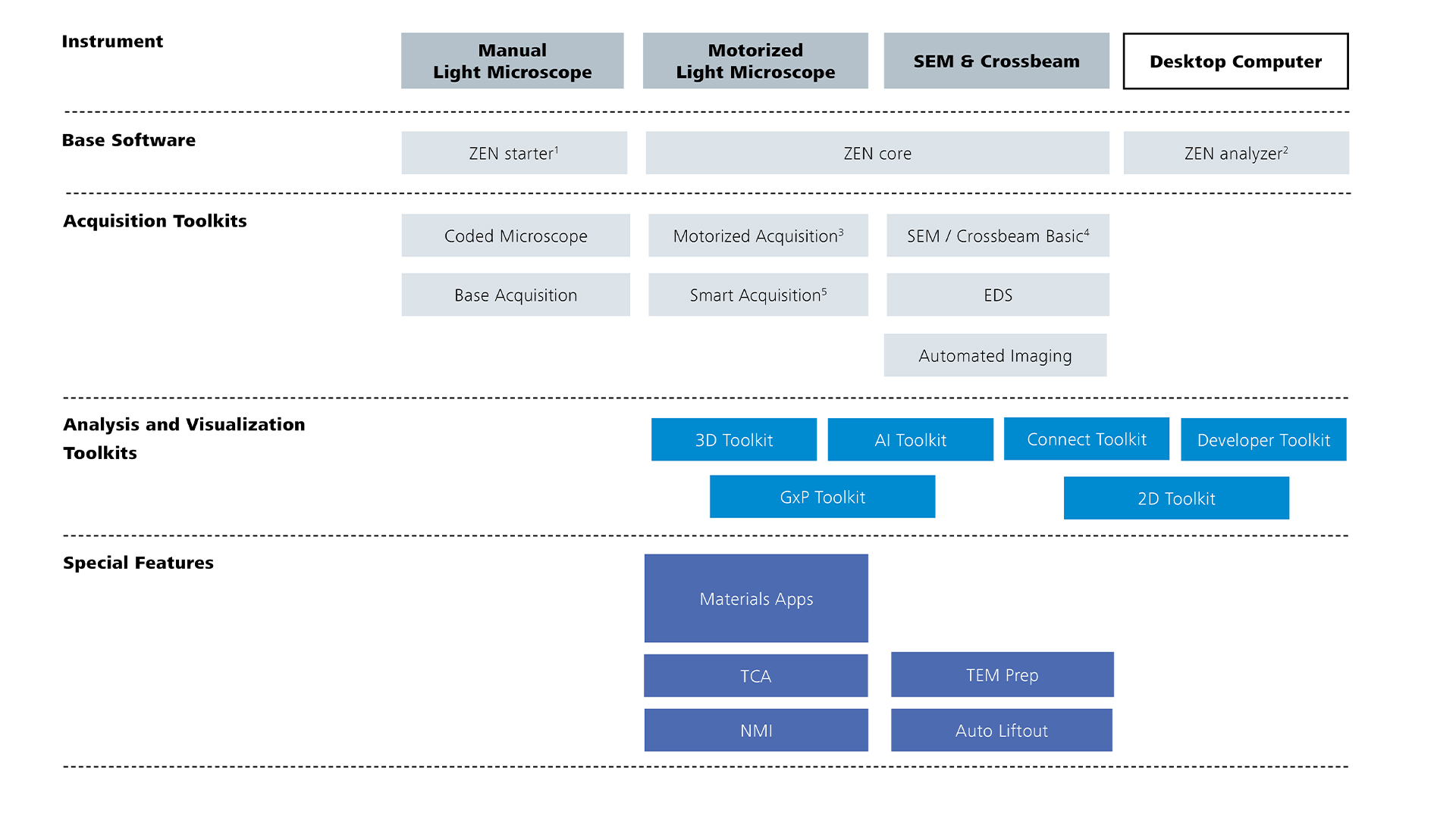
Toolkits
1 Only extensible with Coded Microscope & Based Acquisition
2 No device control and acquisition features
3 Two variants available: with tiling for a motorized stage and without tiling for a manual stage
4 Mandatory for SEM & Crossbeam
5 Requires Motorized Acquisition
1 Only extensible with Coded Microscope & Based Acquisition
2 No device control and acquisition features
3 Two variants available: with tiling for a motorized stage and without tiling for a manual stage
4 Mandatory for SEM & Crossbeam
5 Requires Motorized Acquisition
Discover What’s in For You
The demands of microscopists are not static over time: tasks change, and requirements expand with the progress in research or the need to optimize failure analysis. The ZEN software suite is therefore continuously evolving. Users of ZEN core will benefit from this continuous development now and in the future. Take advantage of updates in the functionalities and enjoy working with new features.
ZEN Toolkits
Base
Perform basic image acquisition of multi-channel fluorescence and time-lapse experiments with manual, coded, and motorized microscopes.
Motorized
Automatically acquire images at different focus positions. Combine them to create an image with a greater depth of field. Record high-resolved images of large samples by automatically scanning pre-defined areas.
Smart Acquisition
Automatically identify positions in overview images and create detail scans.
Coded Microscope Read out of coded microscope components.
EDS Integrated control for the analysis of chemical composition with energy dispersive X-ray spectroscopy.
Automated Imaging
Acquisition of large EM tiled images on ZEISS FE-SEMs via defined imaging protocols.
2D
Employ extended image processing functions and perform automatic 2D image analysis guided by an intuitive software wizard.
3D
Employ extended image processing functions, visualize 3D image stacks with advanced rendering tools, and perform 3D image analysis guided by an intuitive software wizard.
AI
Segment, classify, and denoise images based on machine learning algorithms accessible through a dedicated user interface for training AI models. Training interfaces are included.
Connect
Acquire and correlate images from different instruments such as light and electron microscopes, with a sample-centric workspace and a dedicated file management system. Perform workflows in 2D and 3D, optionally simplified by using L-marker calibration.
Developer
Customize and automate image acquisition, processing, and analysis with a dedicated Python script editor for recording, debugging, and code completion.
GxP
Maintain GxP compliance for your images, tables, and reports when working in regulated environments such as pharmaceutical companies. Ensure traceability and accountability (precondition for 21 CFR Part 11 compliance).
Materials Apps
Perform layer thickness measurements and analyses of grain size, cast iron or multiphases. Compare diagrams with standard charts. Obtain AI-ready results and optionally perform image segmentation or object classification later using the AI Toolkit.
NMI
Perform automated imaging, classification and reporting of non-metallic inclusions in steel.
TCA
Perform automated identification and classification of particles compliant to cleanliness standards.
TEM Prep
Prepare lamellae for TEM investigations on ZEISS Crossbeam using an automated workflow.
Auto Liftout
Automatically lift out and attach readily prepared lamellae to a TEM grid.
ZEN core for Electron Microscopes
Third-party Content Blocked
The video player is blocked due to your cookie preferences. To change the settings and play the video, please click the button below and consent to use of "Functional" tracking technologies.
More than Just the Sum of Its Parts
Benefit from this operational software that is more than just a system control: ZEN core for EM not only simplifies standard SEM workflows but also opens the door for connected microscopy. It links to functionalities such as correlative microscopy, post-processing analyses, reporting, or data management.
Electron microscopists can easily perform standard workflows on every ZEISS SEM and FIB-SEM, from EVO to Sigma to GeminiSEM and Crossbeam.
How it works
Navigation Workspace
ZEN core presents a unique concept featuring a central Navigation Workspace and adaptable workbenches, allowing for easy navigation and intuitive operation.
To understand its capabilities, imagine using a satellite that provides a comprehensive overview of the entire planet Earth while also allowing you to zoom into the image and finally identify the door of your house. Similarly, the Navigation Workspace guides all your workflows. Just follow the tools in the top navigation. Easily find all essential EM control parameters. Gain an overview of the entire sample holder and all your samples easily, collect the acquired data in an image gallery while simultaneously controlling your live image.
Workbenches
ZEN core resembles a real-life workshop: Functions are grouped into workbenches and each workbench offers a dedicated set of tools. The image (“the workpiece”) is taken from workbench to workbench to apply the required tools. You can add, remove and rearrange workbenches and tools as needed for your task.
Uniquely, ZEN core offers the ability to customize the UI to the workflows you need by creating and saving workbenches. Once there, even beginners or users spending little time on the instrument will benefit from just having to open the predefined workbenches and start working on the instrument immediately.
Image acquisition, analytics, and multi-modal, multi-scale workflows can be run by workbenches, such as Sample setup, Acquisition, EDS Processing or Reports.
Integrating Image Acquisition and EDS Analysis
Combining state-of-the-art FE-SEM and EDS technology from ZEISS and Oxford Instruments not only streamlines your EM workflows but also grants unparalleled quality of results. Acquire SEM imagery and rapidly analyze chemical composition without having to switch computers, monitors, keyboards, or software.
Now, a single interface with EDS acquisition and processing workbenches lets you collect analytical data quickly and easily. Analyze a specific ROI. Visualize the elemental composition over the entire field of view with EDS mapping. Process your data afterwards working on the tool or remotely as the Processing Workbench is hardware independent.
Third-party Content Blocked
The video player is blocked due to your cookie preferences. To change the settings and play the video, please click the button below and consent to use of "Functional" tracking technologies.
Watch this video to understand how you will benefit from the Navigation Workspace: Follow the mouse to understand how you can use the image of the sample holder to navigate to your ROI and start image acquisition.
Comparing Segmentation Methods for Particle Analysis of Lithium Ion Battery Materials Using ZEN core
Author: Tim Schubert, Aalen University, Germany
Accurate particle size measurement is vital in materials research, because it affects material properties. This study examines methods for analyzing particle size distributions and geometric features using light optical and scanning electron microscopy (SEM). It tackles the challenges of segmenting individual particles by comparing three techniques: global thresholding, machine learning, and deep-neural-network instance segmentation in lithium-ion battery anodes.
Comparison of three segmentation methods
Analysis of particle sizes with instance segmentation
Segmentation of individual particles with a trained model
Downloads
ZEISS ZEN core
Software Suite for Connected Microscopy in Material Laboratories
As digitalization advances in microscopy, so do the complexities of cybersecurity. ZEISS Microscopy is committed to proactively securing our technologies and protecting our customers. Our Cybersecurity and Data Privacy Governance Program goes beyond traditional security—it also encompasses Responsible AI and Open Source Software (FOSS) governance.