

Untersuchung
Visualisierung und präzise Messung dichter Katarakte


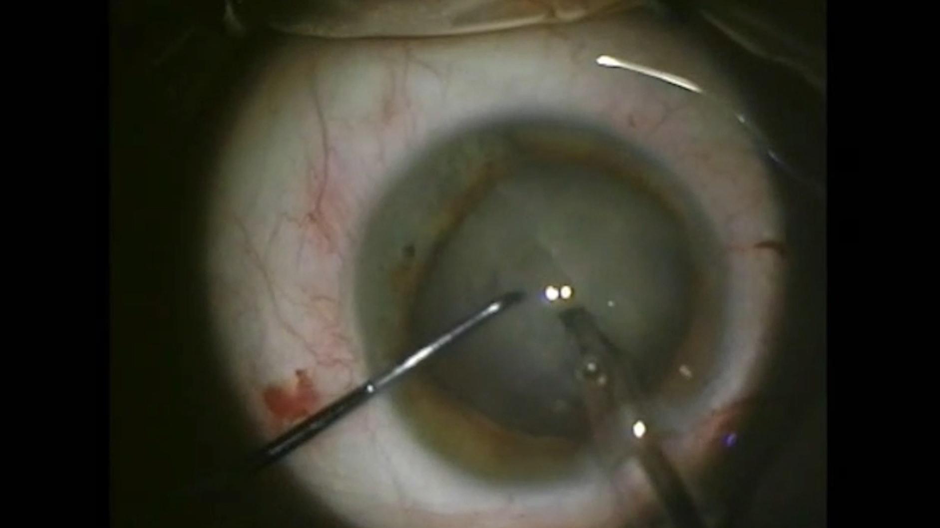
64-jährige Frau mit dichter weißer intumeszenter Katarakt
Screening von Netzhauterkrankungen in der Kataraktchirurgie


ZEISS IOLMaster 700
ZEISS IOLMaster 700 optimiert die Effizienz der Arbeitsabläufe auch im Fall von dichten Katarakten:
- Bis zu 99 % Kataraktdurchdringung, was die Notwendigkeit von Ultraschalluntersuchungen um 92 % reduziert
- Messdauer < 45 Sekunden für beide Augen7
- Der einzigartige Fixation Check lässt Sie Makulaerkrankungen direkt in Ihrem Routine-Workflow erkennen8


ZEISS CIRRUS 6000
Mit ZEISS CIRRUS 6000 kann eine Kataraktpraxis verborgene Pathologien aufdecken und geeignete OP-Kandidaten zum richtigen Zeitpunkt erkennen. Das optimiert die Effizienz Ihrer Arbeitsabläufe in der Praxis.
- 100.000 A-Scans pro Sekunde und ein größeres Sehfeld von bis zu 12 mm – in einem einzigen Scandurchlauf
- Umfangreiche Datenprotokolle mit hoher Detaildichte und verschiedene Bildwinkel bilden die beste Grundlage für exakte diagnostische Ergebnisse
- Zeigt den objektiven Zustand der Makula
- Einfacheres Erwartungsmanagement hinsichtlich des postoperativen Sehvermögens des Patienten


-
1
S. Garg, Surgeons meet challenges of removing rock-hard cataracts, Ocular Surgery News 2018, https://www.healio.com/news/ophthalmology/20181010/surgeons-meet-challenges-of-removing-rockhard-cataracts.
-
2
U. Devgan, Dense brunescent cataracts present surgical challenges, Ocular Surgery News 2011, https://www.healio.com/news/ophthalmology/20120331/dense-brunescent-cataracts-present-surgical-challenges.
-
3
A. Brissette, OCT Is Indispensable for Pre-op Cataract Evaluation, Opthalmology Management 2019, https://www.ophthalmologymanagement.com/newsletters/insights-into-integrated-diagnostic-imaging/may-2019.
-
4
Hirnschall N et al. Macular disease detection with a swept-source optical coherence tomography-based biometry device in patients scheduled for cataract surgery. J Cataract Refract Surg April 2016; 42(4): 530–6.
-
5
Hirnschall N et al. Enhanced Penetration for Axial Length Measurement of Eyes with Dense Cataracts Using Swept Source Optical Coherence Tomography: A Consecutive Observational Study. Ophthalmol Ther 2018; 7: 119–124.
-
6
R. Varsits, N. Hirnschall, B. Doeller, O. Findl; Increasing the number of successful axial eye length measurements using swept-source optical coherence tomography technology compared to conventional optical biometry; präsentiert auf der ESCRS 2016.
-
7
Abhängig von der Augenerkrankung und der Erfahrung des Anwenders.
-
8
ZEISS IOLMaster 700 wurde ausdrücklich nicht für die Diagnostik konzipiert. Ergebnisse müssen verifiziert und Pathologien mit einer speziellen OCT der Netzhaut diagnostiziert werden.
-
9
M. Colvard, Phacoemulsification of the rock hard cataract, Eyeworld 2012, https://www.eyeworld.org/article-phacoemulsification-of-the-rock-hard.
-
10
http://corporate.ewreplay.org/?v=6122663037001.
-
11
R. J. Olson, MD, Salt Lake City, Review of Ophtalmology, publiziert am 15. Januar 2005, Demystifying Dysphotopsia.