On-Demand-Webinar
Virtuelle Gehirnbiopsien: Vergleich der intraoperativen konfokalen Endomikroskopie (CLE) mit der konventionellen Histopathologie
Aufgezeichnet anlässlich des 2. ZEISS Clinical Symposium zu ZEISS CONVIVO
23 Juni 2021
· 20 Min. Lesedauer
![]({"xsmall":"https://www.zeiss.com/content/dam/med-hcp/reference-master/myzeiss/peer-insights/free/pathology/509-neurosurgical-workflow-for-patients-with-brain-tumors/jennifer-eschbacher.jpg/_jcr_content/renditions/original.image_file.100.100.0,0,350,350.file/jennifer-eschbacher.jpg","small":"https://www.zeiss.com/content/dam/med-hcp/reference-master/myzeiss/peer-insights/free/pathology/509-neurosurgical-workflow-for-patients-with-brain-tumors/jennifer-eschbacher.jpg/_jcr_content/renditions/original.image_file.350.350.0,0,350,350.file/jennifer-eschbacher.jpg","medium":"https://www.zeiss.com/content/dam/med-hcp/reference-master/myzeiss/peer-insights/free/pathology/509-neurosurgical-workflow-for-patients-with-brain-tumors/jennifer-eschbacher.jpg/_jcr_content/renditions/original.image_file.350.350.0,0,350,350.file/jennifer-eschbacher.jpg","large":"https://www.zeiss.com/content/dam/med-hcp/reference-master/myzeiss/peer-insights/free/pathology/509-neurosurgical-workflow-for-patients-with-brain-tumors/jennifer-eschbacher.jpg/_jcr_content/renditions/original.image_file.350.350.0,0,350,350.file/jennifer-eschbacher.jpg","xlarge":"https://www.zeiss.com/content/dam/med-hcp/reference-master/myzeiss/peer-insights/free/pathology/509-neurosurgical-workflow-for-patients-with-brain-tumors/jennifer-eschbacher.jpg/_jcr_content/renditions/original.image_file.350.350.0,0,350,350.file/jennifer-eschbacher.jpg","xxlarge":"https://www.zeiss.com/content/dam/med-hcp/reference-master/myzeiss/peer-insights/free/pathology/509-neurosurgical-workflow-for-patients-with-brain-tumors/jennifer-eschbacher.jpg/_jcr_content/renditions/original.image_file.350.350.0,0,350,350.file/jennifer-eschbacher.jpg","max":"https://www.zeiss.com/content/dam/med-hcp/reference-master/myzeiss/peer-insights/free/pathology/509-neurosurgical-workflow-for-patients-with-brain-tumors/jennifer-eschbacher.jpg/_jcr_content/renditions/original.image_file.350.350.0,0,350,350.file/jennifer-eschbacher.jpg"})
Autor
Jennifer Eschbacher, MD
Neuropathologin am Barrow Neurological Institute Phoenix, Arizona, USA
ZUSAMMENFASSUNG
Peer-to-Peer-Erfahrungsaustausch zur Korrelation von intraoperativen CLE-Bildern und konventioneller Histopathologie
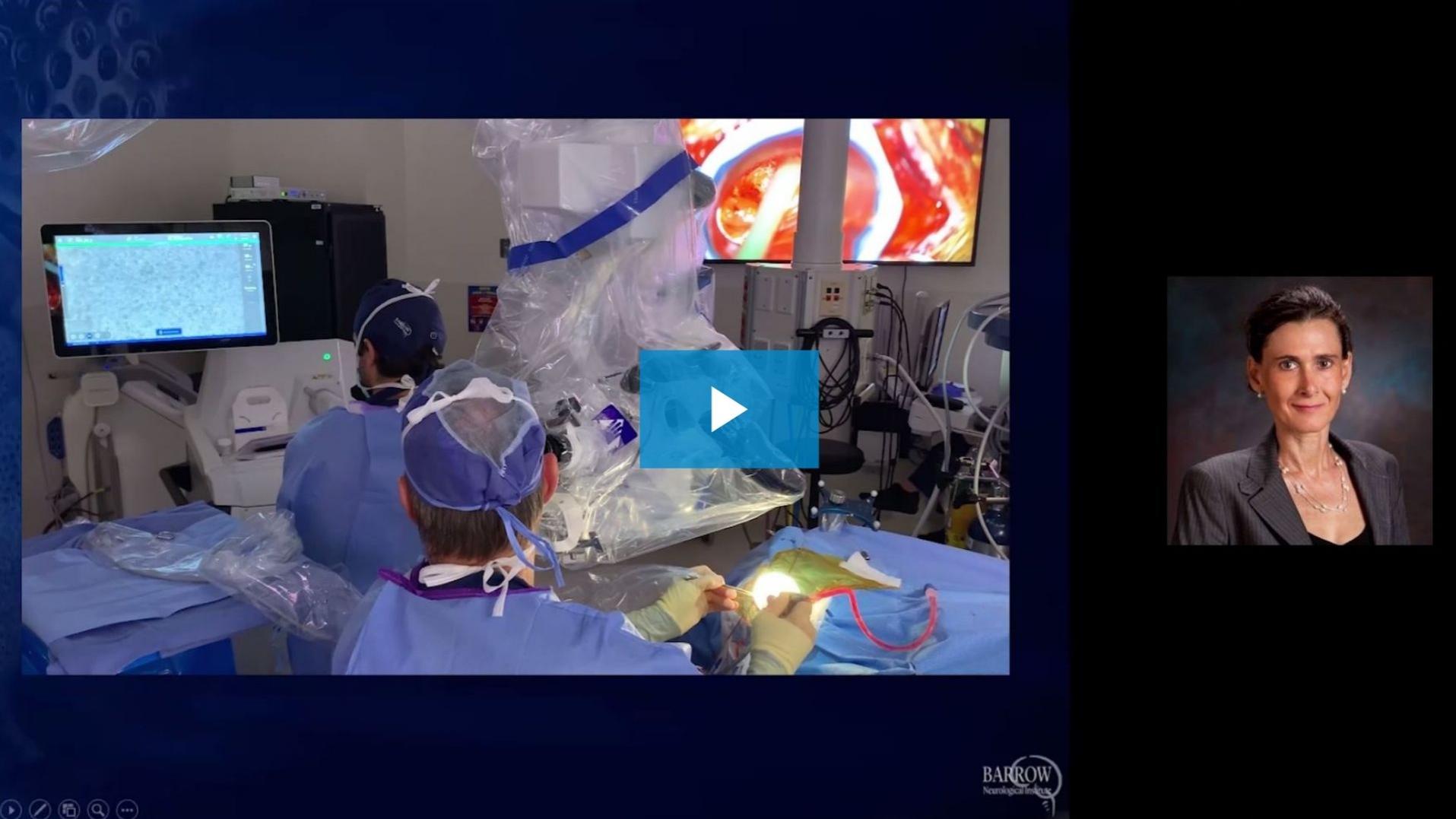
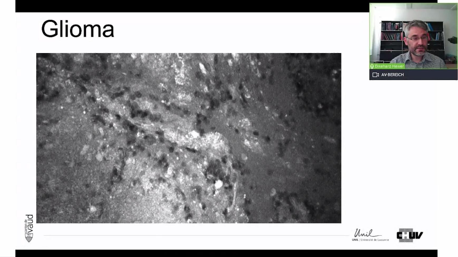
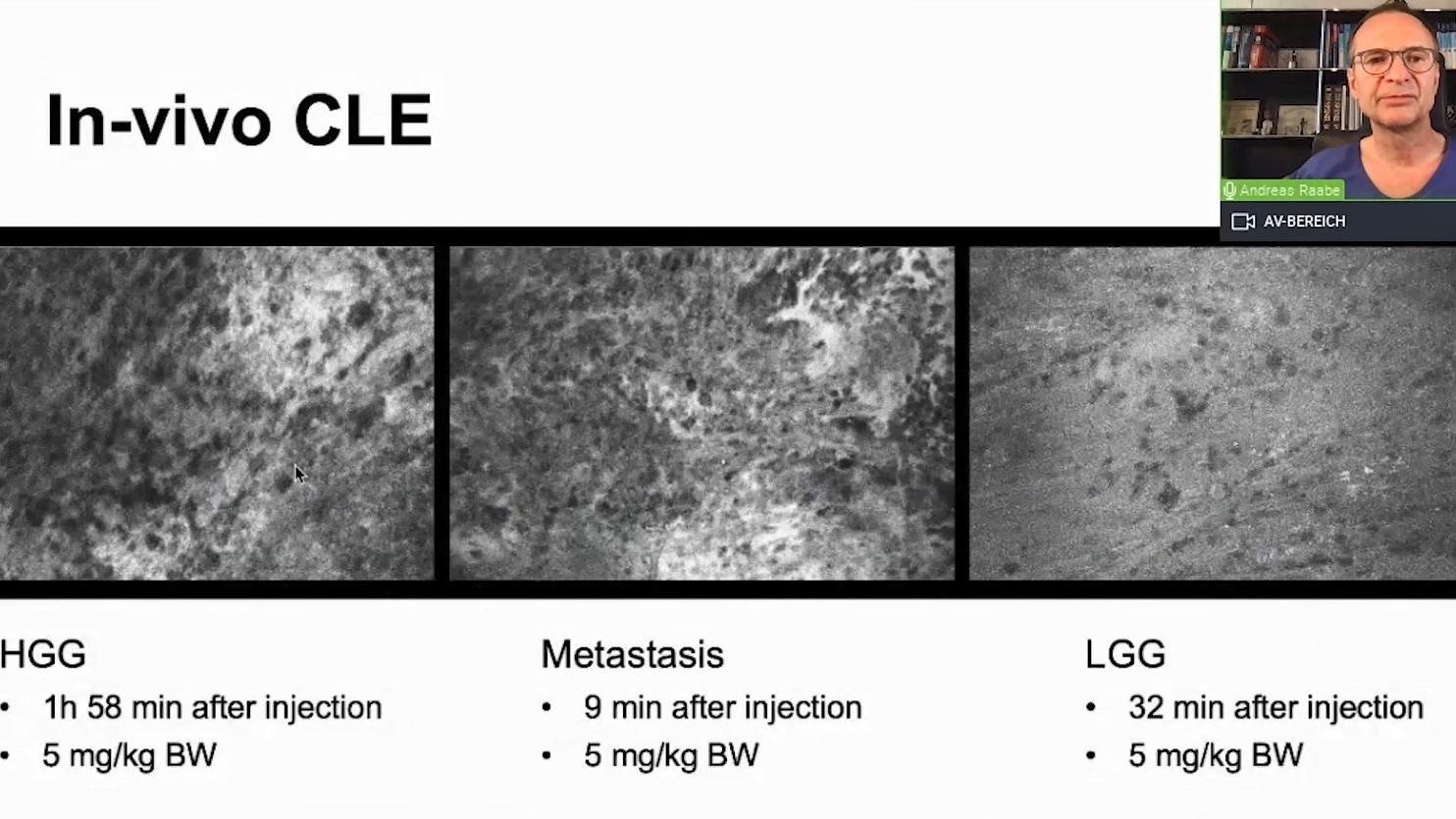
Jennifer Eschbacher, MD, beschreibt ihre Erfahrung mit virtuellen Hirntumorbiopsien auf Zellebene mithilfe konfokaler Endomikroskopie (CLE). In ihrer Präsentation spricht sie über die Ergebnisse der laufenden CLE-Studie. Darüber hinaus gibt sie Einblicke in ein aufgezeichnetes Fallbeispiel einer In-vivo-Pathologie, die sie remote aus dem Pathologielabor verfolgt hat, und demonstriert die Interaktion zwischen Neurochirurgen und Pathologen.
![]({"xsmall":"https://www.zeiss.com/content/dam/med-hcp/reference-master/myzeiss/peer-insights/free/pathology/642-correlation-of-intraoperative-cle-images-and-conventional-histopathology/642--correlation-of-intraoperative-cle-images-and-conventional-h.jpg/_jcr_content/renditions/original.image_file.100.56.0,2,1916,1079.file/642--correlation-of-intraoperative-cle-images-and-conventional-h.jpg","small":"https://www.zeiss.com/content/dam/med-hcp/reference-master/myzeiss/peer-insights/free/pathology/642-correlation-of-intraoperative-cle-images-and-conventional-histopathology/642--correlation-of-intraoperative-cle-images-and-conventional-h.jpg/_jcr_content/renditions/original.image_file.360.203.0,2,1916,1079.file/642--correlation-of-intraoperative-cle-images-and-conventional-h.jpg","medium":"https://www.zeiss.com/content/dam/med-hcp/reference-master/myzeiss/peer-insights/free/pathology/642-correlation-of-intraoperative-cle-images-and-conventional-histopathology/642--correlation-of-intraoperative-cle-images-and-conventional-h.jpg/_jcr_content/renditions/original.image_file.768.432.0,2,1916,1079.file/642--correlation-of-intraoperative-cle-images-and-conventional-h.jpg","large":"https://www.zeiss.com/content/dam/med-hcp/reference-master/myzeiss/peer-insights/free/pathology/642-correlation-of-intraoperative-cle-images-and-conventional-histopathology/642--correlation-of-intraoperative-cle-images-and-conventional-h.jpg/_jcr_content/renditions/original.image_file.1024.576.0,2,1916,1079.file/642--correlation-of-intraoperative-cle-images-and-conventional-h.jpg","xlarge":"https://www.zeiss.com/content/dam/med-hcp/reference-master/myzeiss/peer-insights/free/pathology/642-correlation-of-intraoperative-cle-images-and-conventional-histopathology/642--correlation-of-intraoperative-cle-images-and-conventional-h.jpg/_jcr_content/renditions/original.image_file.1280.720.0,2,1916,1079.file/642--correlation-of-intraoperative-cle-images-and-conventional-h.jpg","xxlarge":"https://www.zeiss.com/content/dam/med-hcp/reference-master/myzeiss/peer-insights/free/pathology/642-correlation-of-intraoperative-cle-images-and-conventional-histopathology/642--correlation-of-intraoperative-cle-images-and-conventional-h.jpg/_jcr_content/renditions/original.image_file.1440.810.0,2,1916,1079.file/642--correlation-of-intraoperative-cle-images-and-conventional-h.jpg","max":"https://www.zeiss.com/content/dam/med-hcp/reference-master/myzeiss/peer-insights/free/pathology/642-correlation-of-intraoperative-cle-images-and-conventional-histopathology/642--correlation-of-intraoperative-cle-images-and-conventional-h.jpg/_jcr_content/renditions/original.image_file.1915.1077.0,2,1916,1079.file/642--correlation-of-intraoperative-cle-images-and-conventional-h.jpg"})