On-Demand-Webinar
Erfahrungen mit der konfokalen Endomikroskopie bei der Resektion von ZNS-Tumoren
Webinar der ZEISS Neuro Week 2020 (Aufzeichnung)
16 Dezember 2020
· 18 Min. Videodauer
![]({"xsmall":"https://www.zeiss.com/content/dam/med-hcp/reference-master/myzeiss/peer-insights/free/pathology/512-ex-vivo-and-in-vivo-fluorescein-guided-confocal-endomicroscopy/francesco-acerbi.jpg/_jcr_content/renditions/original.image_file.100.100.0,190,1355,1545.file/francesco-acerbi.jpg","small":"https://www.zeiss.com/content/dam/med-hcp/reference-master/myzeiss/peer-insights/free/pathology/512-ex-vivo-and-in-vivo-fluorescein-guided-confocal-endomicroscopy/francesco-acerbi.jpg/_jcr_content/renditions/original.image_file.360.360.0,190,1355,1545.file/francesco-acerbi.jpg","medium":"https://www.zeiss.com/content/dam/med-hcp/reference-master/myzeiss/peer-insights/free/pathology/512-ex-vivo-and-in-vivo-fluorescein-guided-confocal-endomicroscopy/francesco-acerbi.jpg/_jcr_content/renditions/original.image_file.768.768.0,190,1355,1545.file/francesco-acerbi.jpg","large":"https://www.zeiss.com/content/dam/med-hcp/reference-master/myzeiss/peer-insights/free/pathology/512-ex-vivo-and-in-vivo-fluorescein-guided-confocal-endomicroscopy/francesco-acerbi.jpg/_jcr_content/renditions/original.image_file.1024.1024.0,190,1355,1545.file/francesco-acerbi.jpg","xlarge":"https://www.zeiss.com/content/dam/med-hcp/reference-master/myzeiss/peer-insights/free/pathology/512-ex-vivo-and-in-vivo-fluorescein-guided-confocal-endomicroscopy/francesco-acerbi.jpg/_jcr_content/renditions/original.image_file.1280.1280.0,190,1355,1545.file/francesco-acerbi.jpg","xxlarge":"https://www.zeiss.com/content/dam/med-hcp/reference-master/myzeiss/peer-insights/free/pathology/512-ex-vivo-and-in-vivo-fluorescein-guided-confocal-endomicroscopy/francesco-acerbi.jpg/_jcr_content/renditions/original.image_file.1355.1355.0,190,1355,1545.file/francesco-acerbi.jpg","max":"https://www.zeiss.com/content/dam/med-hcp/reference-master/myzeiss/peer-insights/free/pathology/512-ex-vivo-and-in-vivo-fluorescein-guided-confocal-endomicroscopy/francesco-acerbi.jpg/_jcr_content/renditions/original.image_file.1355.1355.0,190,1355,1545.file/francesco-acerbi.jpg"})
Autor
Dr. Francesco Acerbi, PhD
Fondazione IRCCS Istituto Neurologico Carlo Besta, Mailand, Italien
ZUSAMMENFASSUNG
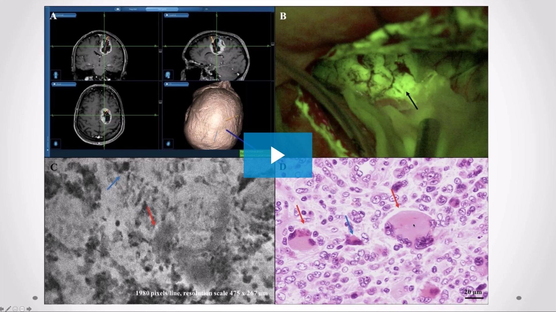
Fluoreszenzgestützte konfokale Endomikroskopie ex vivo und in vivo bei der Resektion von ZNS-Tumoren: Erfahrungen im Istituto Neurologico Carlo Besta
In diesem aufgezeichneten Webinar berichtet Dr. Francesco Acerbi, PhD, von seinen klinischen Erfahrungen mit der fluoreszenzgeführten konfokalen Endomikroskopie bei der Entfernung von ZNS-Tumoren. Er präsentiert Ergebnisse einer Ex-vivo-Studie, in der die diagnostischen und morphologischen Befunde des Tumorgewebes mittels konfokaler Lasermikroskopie mit den Befunden aus der Gefrierschnitt- und der Standardhistologie verglichen werden. Außerdem erörtert er das Potenzial der konfokalen Endomikroskopie für die Verbesserung des Resektionsumfangs (EOR).
![]({"xsmall":"https://www.zeiss.com/content/dam/med-hcp/reference-master/myzeiss/peer-insights/free/pathology/512-ex-vivo-and-in-vivo-fluorescein-guided-confocal-endomicroscopy/512-ex-vivo-and-in-vivo-fluorescein-guided-confocal-endomicrosco_preview.jpg/_jcr_content/renditions/original.image_file.100.56.5,6,1915,1080.file/512-ex-vivo-and-in-vivo-fluorescein-guided-confocal-endomicrosco_preview.jpg","small":"https://www.zeiss.com/content/dam/med-hcp/reference-master/myzeiss/peer-insights/free/pathology/512-ex-vivo-and-in-vivo-fluorescein-guided-confocal-endomicroscopy/512-ex-vivo-and-in-vivo-fluorescein-guided-confocal-endomicrosco_preview.jpg/_jcr_content/renditions/original.image_file.360.203.5,6,1915,1080.file/512-ex-vivo-and-in-vivo-fluorescein-guided-confocal-endomicrosco_preview.jpg","medium":"https://www.zeiss.com/content/dam/med-hcp/reference-master/myzeiss/peer-insights/free/pathology/512-ex-vivo-and-in-vivo-fluorescein-guided-confocal-endomicroscopy/512-ex-vivo-and-in-vivo-fluorescein-guided-confocal-endomicrosco_preview.jpg/_jcr_content/renditions/original.image_file.768.432.5,6,1915,1080.file/512-ex-vivo-and-in-vivo-fluorescein-guided-confocal-endomicrosco_preview.jpg","large":"https://www.zeiss.com/content/dam/med-hcp/reference-master/myzeiss/peer-insights/free/pathology/512-ex-vivo-and-in-vivo-fluorescein-guided-confocal-endomicroscopy/512-ex-vivo-and-in-vivo-fluorescein-guided-confocal-endomicrosco_preview.jpg/_jcr_content/renditions/original.image_file.1024.576.5,6,1915,1080.file/512-ex-vivo-and-in-vivo-fluorescein-guided-confocal-endomicrosco_preview.jpg","xlarge":"https://www.zeiss.com/content/dam/med-hcp/reference-master/myzeiss/peer-insights/free/pathology/512-ex-vivo-and-in-vivo-fluorescein-guided-confocal-endomicroscopy/512-ex-vivo-and-in-vivo-fluorescein-guided-confocal-endomicrosco_preview.jpg/_jcr_content/renditions/original.image_file.1280.720.5,6,1915,1080.file/512-ex-vivo-and-in-vivo-fluorescein-guided-confocal-endomicrosco_preview.jpg","xxlarge":"https://www.zeiss.com/content/dam/med-hcp/reference-master/myzeiss/peer-insights/free/pathology/512-ex-vivo-and-in-vivo-fluorescein-guided-confocal-endomicroscopy/512-ex-vivo-and-in-vivo-fluorescein-guided-confocal-endomicrosco_preview.jpg/_jcr_content/renditions/original.image_file.1440.810.5,6,1915,1080.file/512-ex-vivo-and-in-vivo-fluorescein-guided-confocal-endomicrosco_preview.jpg","max":"https://www.zeiss.com/content/dam/med-hcp/reference-master/myzeiss/peer-insights/free/pathology/512-ex-vivo-and-in-vivo-fluorescein-guided-confocal-endomicroscopy/512-ex-vivo-and-in-vivo-fluorescein-guided-confocal-endomicrosco_preview.jpg/_jcr_content/renditions/original.image_file.1910.1074.5,6,1915,1080.file/512-ex-vivo-and-in-vivo-fluorescein-guided-confocal-endomicrosco_preview.jpg"})