On-Demand-Webinar
Konfokale Endomikroskopie in der Neurochirurgie
Bei der EANS 2019 aufgezeichneter Vortrag
24 Juli 2019
· 19 Min. Videodauer
![]({"xsmall":"https://www.zeiss.com/content/dam/med-hcp/reference-master/myzeiss/peer-insights/free/pathology/382---confocal-endomicroscopy-in-neurosurgery/francesco-acerb-picture.jpg/_jcr_content/renditions/original.image_file.100.100.0,190,1355,1545.file/francesco-acerb-picture.jpg","small":"https://www.zeiss.com/content/dam/med-hcp/reference-master/myzeiss/peer-insights/free/pathology/382---confocal-endomicroscopy-in-neurosurgery/francesco-acerb-picture.jpg/_jcr_content/renditions/original.image_file.360.360.0,190,1355,1545.file/francesco-acerb-picture.jpg","medium":"https://www.zeiss.com/content/dam/med-hcp/reference-master/myzeiss/peer-insights/free/pathology/382---confocal-endomicroscopy-in-neurosurgery/francesco-acerb-picture.jpg/_jcr_content/renditions/original.image_file.768.768.0,190,1355,1545.file/francesco-acerb-picture.jpg","large":"https://www.zeiss.com/content/dam/med-hcp/reference-master/myzeiss/peer-insights/free/pathology/382---confocal-endomicroscopy-in-neurosurgery/francesco-acerb-picture.jpg/_jcr_content/renditions/original.image_file.1024.1024.0,190,1355,1545.file/francesco-acerb-picture.jpg","xlarge":"https://www.zeiss.com/content/dam/med-hcp/reference-master/myzeiss/peer-insights/free/pathology/382---confocal-endomicroscopy-in-neurosurgery/francesco-acerb-picture.jpg/_jcr_content/renditions/original.image_file.1280.1280.0,190,1355,1545.file/francesco-acerb-picture.jpg","xxlarge":"https://www.zeiss.com/content/dam/med-hcp/reference-master/myzeiss/peer-insights/free/pathology/382---confocal-endomicroscopy-in-neurosurgery/francesco-acerb-picture.jpg/_jcr_content/renditions/original.image_file.1355.1355.0,190,1355,1545.file/francesco-acerb-picture.jpg","max":"https://www.zeiss.com/content/dam/med-hcp/reference-master/myzeiss/peer-insights/free/pathology/382---confocal-endomicroscopy-in-neurosurgery/francesco-acerb-picture.jpg/_jcr_content/renditions/original.image_file.1355.1355.0,190,1355,1545.file/francesco-acerb-picture.jpg"})
Autor
Dr. Francesco Acerbi, PhD
Abteilung Neurochirurgie, Fondazione IRCCS Istituto Neurologico Carlo Besta, Mailand, Italien
ZUSAMMENFASSUNG
Erfahrungen mit konfokaler Lasermikroskopie (ex vivo und in vivo) bei Patienten mit intrakraniellen Tumoren
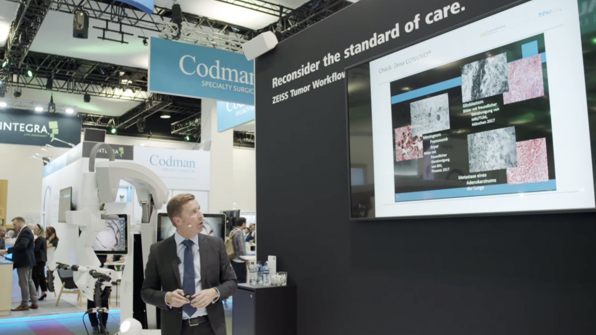
In diesem aufgezeichneten Vortrag berichtet Dr. Francesco Acerbi, PhD, von seinen bisherigen klinischen Erfahrungen mit der konfokalen Endomikroskopie unter Anwendung von Fluorescein-Natrium bei Patienten mit intrakraniellen Tumoren. Präsentiert werden Ex-vivo- und In-Vivo-Fallbeispiele.
![]({"xsmall":"https://www.zeiss.com/content/dam/med-hcp/reference-master/myzeiss/peer-insights/free/pathology/382---confocal-endomicroscopy-in-neurosurgery/confocal-laser-endomicroscopy-for-intracranial-tumors-en-30-160-0384i_video.jpg/_jcr_content/renditions/original.image_file.100.56.file/confocal-laser-endomicroscopy-for-intracranial-tumors-en-30-160-0384i_video.jpg","small":"https://www.zeiss.com/content/dam/med-hcp/reference-master/myzeiss/peer-insights/free/pathology/382---confocal-endomicroscopy-in-neurosurgery/confocal-laser-endomicroscopy-for-intracranial-tumors-en-30-160-0384i_video.jpg/_jcr_content/renditions/original.image_file.360.203.file/confocal-laser-endomicroscopy-for-intracranial-tumors-en-30-160-0384i_video.jpg","medium":"https://www.zeiss.com/content/dam/med-hcp/reference-master/myzeiss/peer-insights/free/pathology/382---confocal-endomicroscopy-in-neurosurgery/confocal-laser-endomicroscopy-for-intracranial-tumors-en-30-160-0384i_video.jpg/_jcr_content/renditions/original.image_file.768.432.file/confocal-laser-endomicroscopy-for-intracranial-tumors-en-30-160-0384i_video.jpg","large":"https://www.zeiss.com/content/dam/med-hcp/reference-master/myzeiss/peer-insights/free/pathology/382---confocal-endomicroscopy-in-neurosurgery/confocal-laser-endomicroscopy-for-intracranial-tumors-en-30-160-0384i_video.jpg/_jcr_content/renditions/original.image_file.1024.576.file/confocal-laser-endomicroscopy-for-intracranial-tumors-en-30-160-0384i_video.jpg","xlarge":"https://www.zeiss.com/content/dam/med-hcp/reference-master/myzeiss/peer-insights/free/pathology/382---confocal-endomicroscopy-in-neurosurgery/confocal-laser-endomicroscopy-for-intracranial-tumors-en-30-160-0384i_video.jpg/_jcr_content/renditions/original.image_file.1280.720.file/confocal-laser-endomicroscopy-for-intracranial-tumors-en-30-160-0384i_video.jpg","xxlarge":"https://www.zeiss.com/content/dam/med-hcp/reference-master/myzeiss/peer-insights/free/pathology/382---confocal-endomicroscopy-in-neurosurgery/confocal-laser-endomicroscopy-for-intracranial-tumors-en-30-160-0384i_video.jpg/_jcr_content/renditions/original.image_file.1440.810.file/confocal-laser-endomicroscopy-for-intracranial-tumors-en-30-160-0384i_video.jpg","max":"https://www.zeiss.com/content/dam/med-hcp/reference-master/myzeiss/peer-insights/free/pathology/382---confocal-endomicroscopy-in-neurosurgery/confocal-laser-endomicroscopy-for-intracranial-tumors-en-30-160-0384i_video.jpg/_jcr_content/renditions/original./confocal-laser-endomicroscopy-for-intracranial-tumors-en-30-160-0384i_video.jpg"})