ZEISS CONVIVO Image Library
for neurosurgery and pathology professionals working with ZEISS In Vivo Pathology Suite
- Gain impressions on ZEISS CONVIVO images of tissue microstructures
- Increase your confidence by comparing with similar cases
- Benefit from case examples for education purposes
Image database to facilitate effective learning of confocal image reading
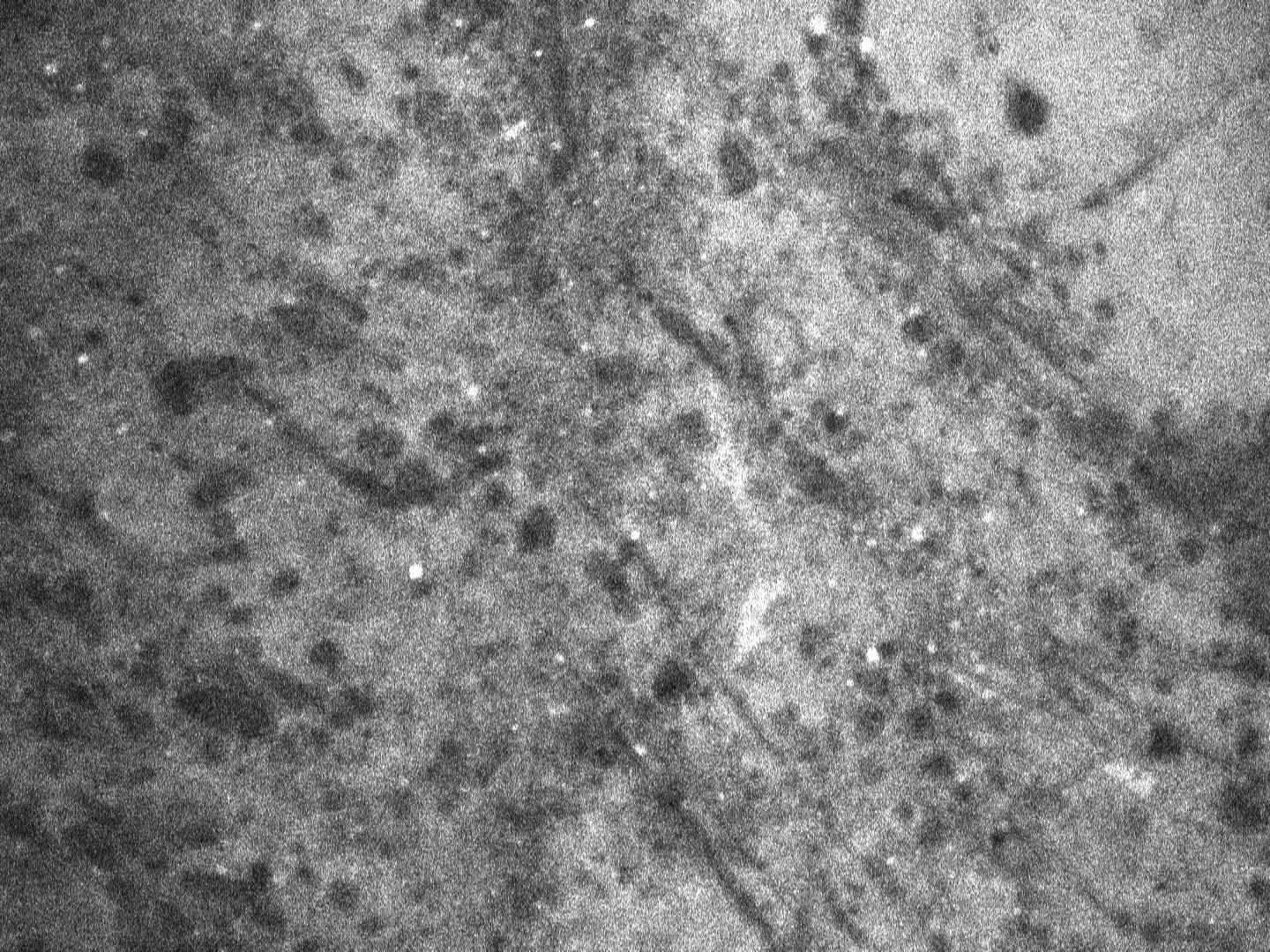
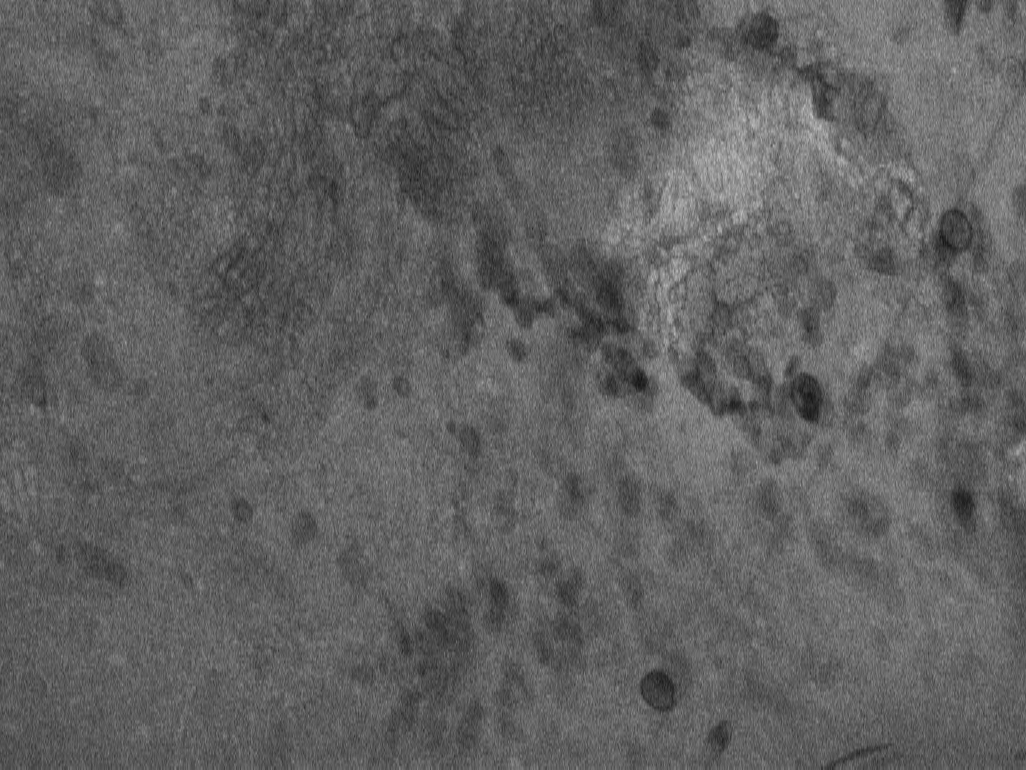
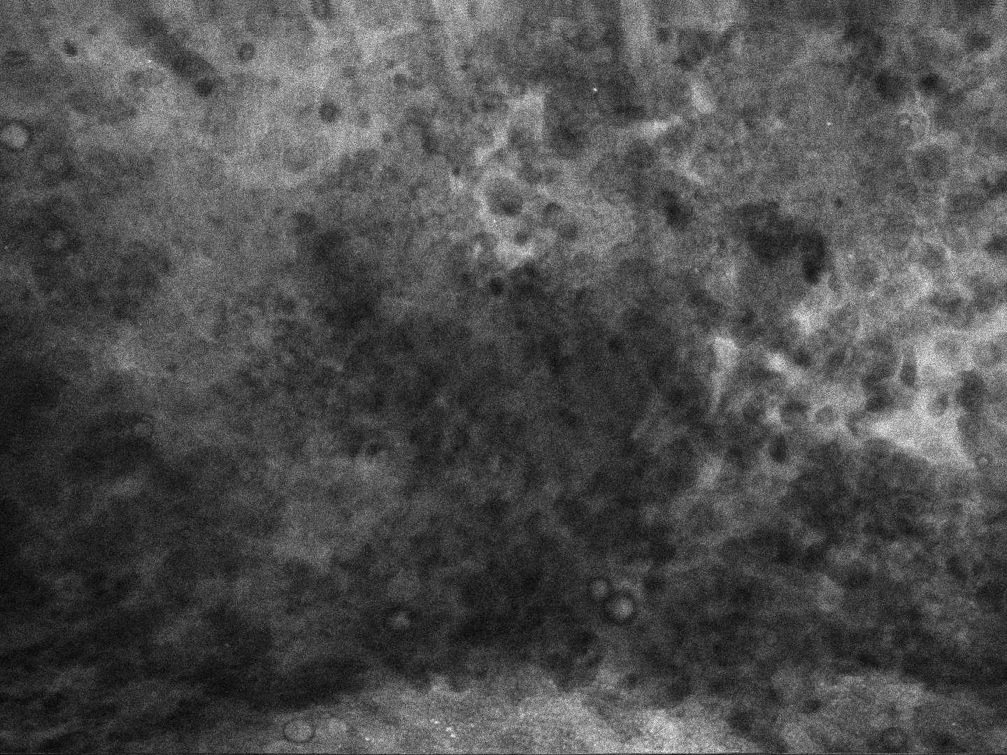
ZEISS CONVIVO Image Library provides selected clinical cases from your peers - showing image examples acquired during procedures performed with CONVIVO® from ZEISS. Explore selected images to get a first impression of how features known from conventional histology can be reproduced with confocal endomicroscopy.1


-
1
ZEISS CONVIVO images are not a substitute for H&E-stains. Actual diagnosis of these cases was based on the standard of care (histopathology / H&E-stain).