Case
Microscope as precision tool – clinical complex case report
15 October 2021
· 15 min read
![]({"xsmall":"https://www.zeiss.com/content/dam/med-hcp/reference-master/myzeiss/peer-insights/free/dentistry/767-peri-implantitis-treatment-with-laser-under-magnification/images/arielsavion-portrait_square.jpg/_jcr_content/renditions/original.image_file.100.100.0,8,476,484.file/arielsavion-portrait_square.jpg","small":"https://www.zeiss.com/content/dam/med-hcp/reference-master/myzeiss/peer-insights/free/dentistry/767-peri-implantitis-treatment-with-laser-under-magnification/images/arielsavion-portrait_square.jpg/_jcr_content/renditions/original.image_file.360.360.0,8,476,484.file/arielsavion-portrait_square.jpg","medium":"https://www.zeiss.com/content/dam/med-hcp/reference-master/myzeiss/peer-insights/free/dentistry/767-peri-implantitis-treatment-with-laser-under-magnification/images/arielsavion-portrait_square.jpg/_jcr_content/renditions/original.image_file.476.476.0,8,476,484.file/arielsavion-portrait_square.jpg","large":"https://www.zeiss.com/content/dam/med-hcp/reference-master/myzeiss/peer-insights/free/dentistry/767-peri-implantitis-treatment-with-laser-under-magnification/images/arielsavion-portrait_square.jpg/_jcr_content/renditions/original.image_file.476.476.0,8,476,484.file/arielsavion-portrait_square.jpg","xlarge":"https://www.zeiss.com/content/dam/med-hcp/reference-master/myzeiss/peer-insights/free/dentistry/767-peri-implantitis-treatment-with-laser-under-magnification/images/arielsavion-portrait_square.jpg/_jcr_content/renditions/original.image_file.476.476.0,8,476,484.file/arielsavion-portrait_square.jpg","xxlarge":"https://www.zeiss.com/content/dam/med-hcp/reference-master/myzeiss/peer-insights/free/dentistry/767-peri-implantitis-treatment-with-laser-under-magnification/images/arielsavion-portrait_square.jpg/_jcr_content/renditions/original.image_file.476.476.0,8,476,484.file/arielsavion-portrait_square.jpg","max":"https://www.zeiss.com/content/dam/med-hcp/reference-master/myzeiss/peer-insights/free/dentistry/767-peri-implantitis-treatment-with-laser-under-magnification/images/arielsavion-portrait_square.jpg/_jcr_content/renditions/original.image_file.476.476.0,8,476,484.file/arielsavion-portrait_square.jpg"})
Author
Dr. Ariel Savion, D.M.D, LL.B, M.Sc, I.C.O.I
Rishon Lezion, Israel
Abstract
Peer-to-peer experience sharing on a complex case of rehabilitation in the upper jaw with old porcelain-fused metal crowns in teeth 1.2 - 2.2
This case concerns a 29-year-old patient who complained about unaesthetic and poor dental brilliancy. During the clinical interview, the patient stated that evidence of this can be found in the intra-oral and dento-labial image as well as in the patient’s facial portrait. All images were required to generate a correctly analyzed aesthetic preview.
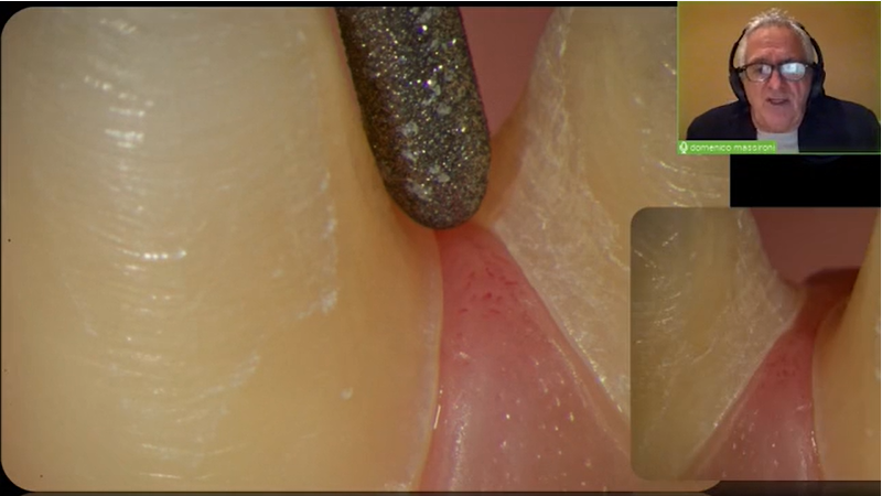
The whole case was treated under the ZEISS EXTARO 300 microscope (ZEISS, Germany) to maximize precision in aesthetic outcome.