Expert discussion
Confocal endomicroscopy for in vivo visualization in brain tumor surgery
11 April 2022
· 10 min watch


Author
Jürgen Schlegel, MD
Department of Neuropathology, Technical University Munich, Germany


Author
Jens Gempt, MD
Department of Neurosurgery, Technical University Munich, Germany


Author
Mark Preul, MD
Department of Neurosurgery, Barrow Neurological Institute, Phoenix, USA
Summary
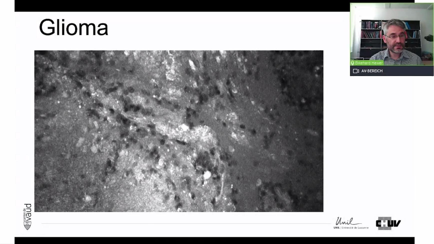
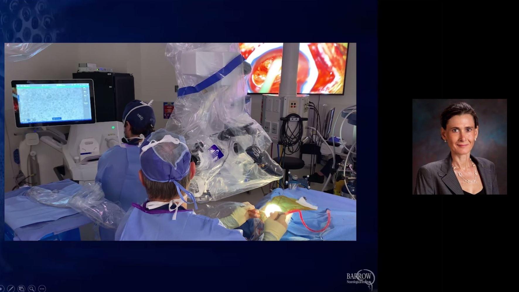
Real-world experience with in vivo tissue visualization using CLE during brain tumor surgery
In this recorded panel discussion, Jürgen Schlegel, MD, Jens Gempt, MD, and Mark C. Preul, MD exchange on the current role of confocal laser endomicroscopy in brain tumor surgery. Based on actual clinical experiences with this technology, the speakers cover topics such as in vivo CLE and ex vivo CLE study results, confocal image acquisition, and the role of different benefits for the multidisciplinary collaboration.