On-demand webinar
In vivo experience using confocal endomicroscopy
Expert presentation and discussion recorded during first CONVIVO Clinical Symposium
18 December 2020
· 33 min watch
![]({"xsmall":"https://www.zeiss.com/content/dam/med-hcp/reference-master/myzeiss/peer-insights/free/pathology/509-neurosurgical-workflow-for-patients-with-brain-tumors/jennifer-eschbacher.jpg/_jcr_content/renditions/original.image_file.100.100.0,0,350,350.file/jennifer-eschbacher.jpg","small":"https://www.zeiss.com/content/dam/med-hcp/reference-master/myzeiss/peer-insights/free/pathology/509-neurosurgical-workflow-for-patients-with-brain-tumors/jennifer-eschbacher.jpg/_jcr_content/renditions/original.image_file.350.350.0,0,350,350.file/jennifer-eschbacher.jpg","medium":"https://www.zeiss.com/content/dam/med-hcp/reference-master/myzeiss/peer-insights/free/pathology/509-neurosurgical-workflow-for-patients-with-brain-tumors/jennifer-eschbacher.jpg/_jcr_content/renditions/original.image_file.350.350.0,0,350,350.file/jennifer-eschbacher.jpg","large":"https://www.zeiss.com/content/dam/med-hcp/reference-master/myzeiss/peer-insights/free/pathology/509-neurosurgical-workflow-for-patients-with-brain-tumors/jennifer-eschbacher.jpg/_jcr_content/renditions/original.image_file.350.350.0,0,350,350.file/jennifer-eschbacher.jpg","xlarge":"https://www.zeiss.com/content/dam/med-hcp/reference-master/myzeiss/peer-insights/free/pathology/509-neurosurgical-workflow-for-patients-with-brain-tumors/jennifer-eschbacher.jpg/_jcr_content/renditions/original.image_file.350.350.0,0,350,350.file/jennifer-eschbacher.jpg","xxlarge":"https://www.zeiss.com/content/dam/med-hcp/reference-master/myzeiss/peer-insights/free/pathology/509-neurosurgical-workflow-for-patients-with-brain-tumors/jennifer-eschbacher.jpg/_jcr_content/renditions/original.image_file.350.350.0,0,350,350.file/jennifer-eschbacher.jpg","max":"https://www.zeiss.com/content/dam/med-hcp/reference-master/myzeiss/peer-insights/free/pathology/509-neurosurgical-workflow-for-patients-with-brain-tumors/jennifer-eschbacher.jpg/_jcr_content/renditions/original.image_file.350.350.0,0,350,350.file/jennifer-eschbacher.jpg"})
Author
Jennifer Eschbacher, MD
Barrow Neurological Institute, Phoenix, Arizona, USA
SUMMARY
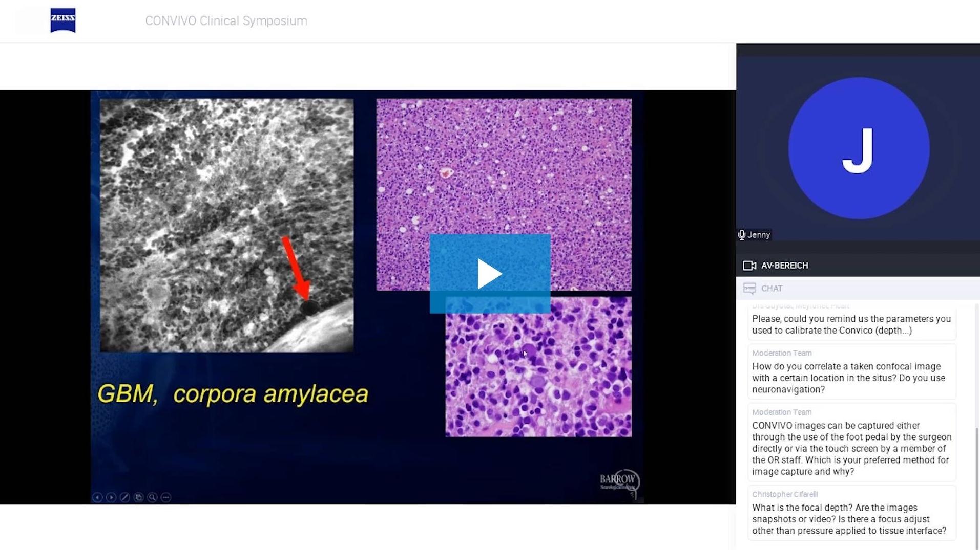
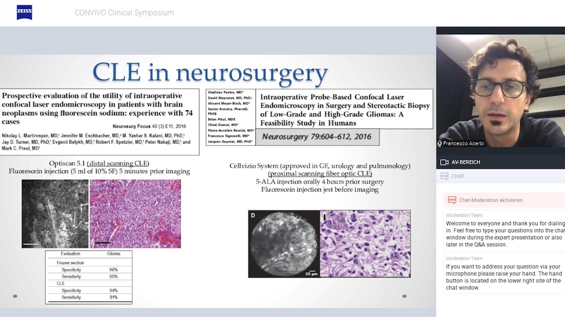
In vivo experience using confocal endomicroscopy at Barrow Neurological Institute, Phoenix – Pathologist perspective
During this recorded webinar, Dr. Jennifer Eschbacher shares her experiences with fluorescein-guided confocal endomicroscopy at Barrow Neurological Institute. In her talk, she discusses limitations of frozen section biopsies and shows the cellularity of confocal and H&E-stained image pairs with the aid of different case examples. Furthermore, she addresses advantages of an in vivo pathology consultation workflow for cross-functional medical teams. Her presentation is followed by a short discussion, in which Dr. Eschbacher answers questions from the audience providing insights as well as practical guidance.
![]({"xsmall":"https://www.zeiss.com/content/dam/med-hcp/reference-master/myzeiss/peer-insights/free/pathology/522-confocal-endomicroscopy-in-vivo-experiences/522-confocal-endomicroscopy-in-vivo-experiences_preview.jpg/_jcr_content/renditions/original.image_file.100.56.3,5,1916,1081.file/522-confocal-endomicroscopy-in-vivo-experiences_preview.jpg","small":"https://www.zeiss.com/content/dam/med-hcp/reference-master/myzeiss/peer-insights/free/pathology/522-confocal-endomicroscopy-in-vivo-experiences/522-confocal-endomicroscopy-in-vivo-experiences_preview.jpg/_jcr_content/renditions/original.image_file.360.203.3,5,1916,1081.file/522-confocal-endomicroscopy-in-vivo-experiences_preview.jpg","medium":"https://www.zeiss.com/content/dam/med-hcp/reference-master/myzeiss/peer-insights/free/pathology/522-confocal-endomicroscopy-in-vivo-experiences/522-confocal-endomicroscopy-in-vivo-experiences_preview.jpg/_jcr_content/renditions/original.image_file.768.432.3,5,1916,1081.file/522-confocal-endomicroscopy-in-vivo-experiences_preview.jpg","large":"https://www.zeiss.com/content/dam/med-hcp/reference-master/myzeiss/peer-insights/free/pathology/522-confocal-endomicroscopy-in-vivo-experiences/522-confocal-endomicroscopy-in-vivo-experiences_preview.jpg/_jcr_content/renditions/original.image_file.1024.576.3,5,1916,1081.file/522-confocal-endomicroscopy-in-vivo-experiences_preview.jpg","xlarge":"https://www.zeiss.com/content/dam/med-hcp/reference-master/myzeiss/peer-insights/free/pathology/522-confocal-endomicroscopy-in-vivo-experiences/522-confocal-endomicroscopy-in-vivo-experiences_preview.jpg/_jcr_content/renditions/original.image_file.1280.720.3,5,1916,1081.file/522-confocal-endomicroscopy-in-vivo-experiences_preview.jpg","xxlarge":"https://www.zeiss.com/content/dam/med-hcp/reference-master/myzeiss/peer-insights/free/pathology/522-confocal-endomicroscopy-in-vivo-experiences/522-confocal-endomicroscopy-in-vivo-experiences_preview.jpg/_jcr_content/renditions/original.image_file.1440.810.3,5,1916,1081.file/522-confocal-endomicroscopy-in-vivo-experiences_preview.jpg","max":"https://www.zeiss.com/content/dam/med-hcp/reference-master/myzeiss/peer-insights/free/pathology/522-confocal-endomicroscopy-in-vivo-experiences/522-confocal-endomicroscopy-in-vivo-experiences_preview.jpg/_jcr_content/renditions/original.image_file.1913.1076.3,5,1916,1081.file/522-confocal-endomicroscopy-in-vivo-experiences_preview.jpg"})