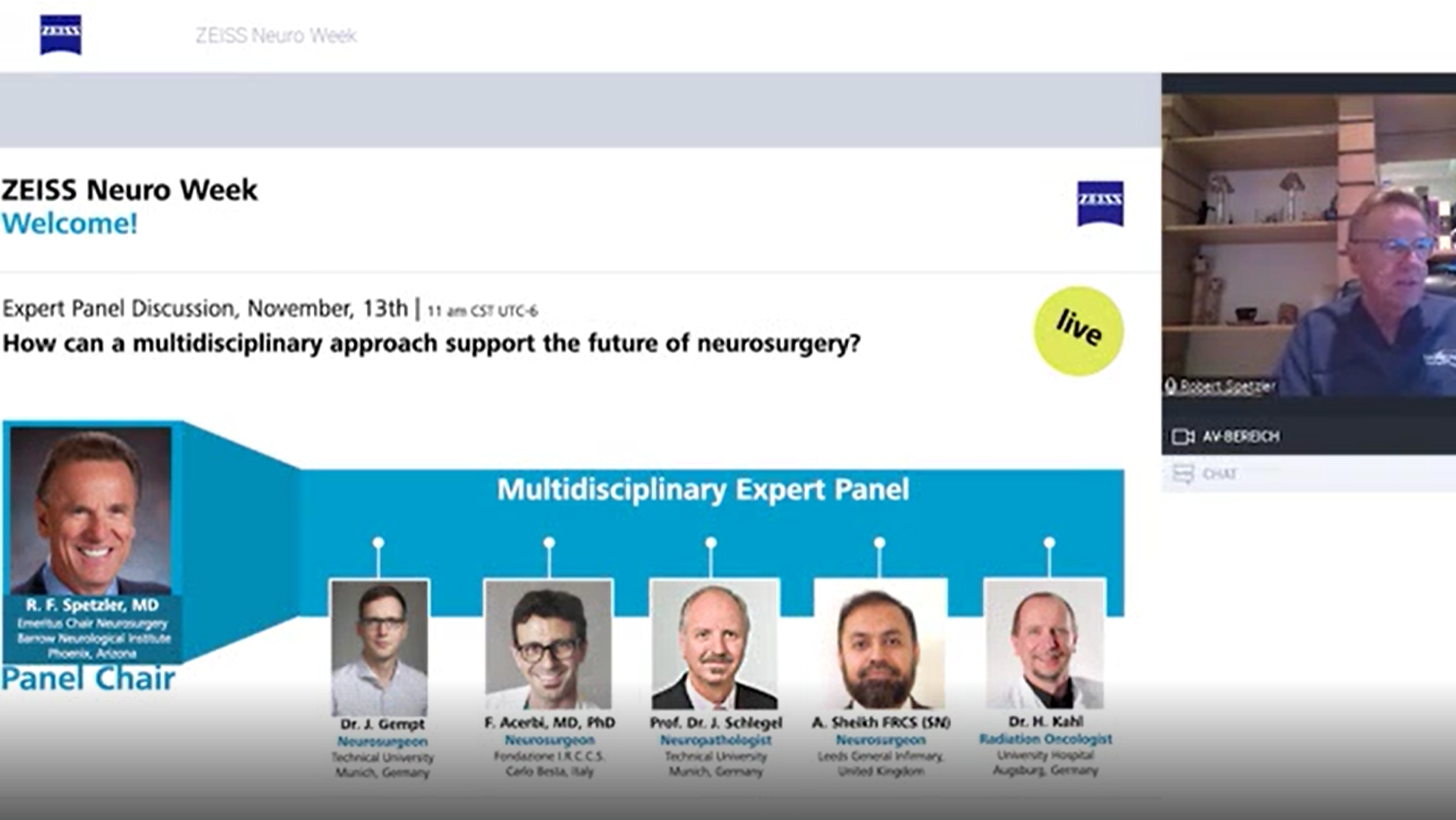
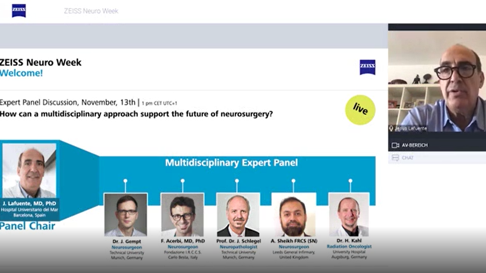
On-demand webinar
Experiences with confocal endomicroscopy during resection of CNS tumors
Webinar recorded during ZEISS Neuro Week 2020
16 December 2020
· 18 min watch
![]({"xsmall":"https://www.zeiss.com/content/dam/med-hcp/reference-master/myzeiss/peer-insights/free/pathology/512-ex-vivo-and-in-vivo-fluorescein-guided-confocal-endomicroscopy/francesco-acerbi.jpg/_jcr_content/renditions/original.image_file.100.100.0,190,1355,1545.file/francesco-acerbi.jpg","small":"https://www.zeiss.com/content/dam/med-hcp/reference-master/myzeiss/peer-insights/free/pathology/512-ex-vivo-and-in-vivo-fluorescein-guided-confocal-endomicroscopy/francesco-acerbi.jpg/_jcr_content/renditions/original.image_file.360.360.0,190,1355,1545.file/francesco-acerbi.jpg","medium":"https://www.zeiss.com/content/dam/med-hcp/reference-master/myzeiss/peer-insights/free/pathology/512-ex-vivo-and-in-vivo-fluorescein-guided-confocal-endomicroscopy/francesco-acerbi.jpg/_jcr_content/renditions/original.image_file.768.768.0,190,1355,1545.file/francesco-acerbi.jpg","large":"https://www.zeiss.com/content/dam/med-hcp/reference-master/myzeiss/peer-insights/free/pathology/512-ex-vivo-and-in-vivo-fluorescein-guided-confocal-endomicroscopy/francesco-acerbi.jpg/_jcr_content/renditions/original.image_file.1024.1024.0,190,1355,1545.file/francesco-acerbi.jpg","xlarge":"https://www.zeiss.com/content/dam/med-hcp/reference-master/myzeiss/peer-insights/free/pathology/512-ex-vivo-and-in-vivo-fluorescein-guided-confocal-endomicroscopy/francesco-acerbi.jpg/_jcr_content/renditions/original.image_file.1280.1280.0,190,1355,1545.file/francesco-acerbi.jpg","xxlarge":"https://www.zeiss.com/content/dam/med-hcp/reference-master/myzeiss/peer-insights/free/pathology/512-ex-vivo-and-in-vivo-fluorescein-guided-confocal-endomicroscopy/francesco-acerbi.jpg/_jcr_content/renditions/original.image_file.1355.1355.0,190,1355,1545.file/francesco-acerbi.jpg","max":"https://www.zeiss.com/content/dam/med-hcp/reference-master/myzeiss/peer-insights/free/pathology/512-ex-vivo-and-in-vivo-fluorescein-guided-confocal-endomicroscopy/francesco-acerbi.jpg/_jcr_content/renditions/original.image_file.1355.1355.0,190,1355,1545.file/francesco-acerbi.jpg"})
Author
Francesco Acerbi, MD, PhD
Foundation IRCCS Istituto Neurologico Besta, Milano, Italy
SUMMARY
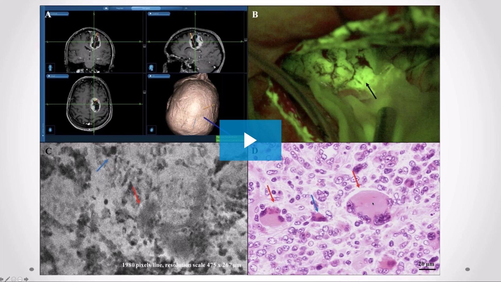
Ex vivo and in vivo fluorescein-guided confocal endomicroscopy during resection of CNS tumors: Experience at Besta Neurological Institute
In this recorded webinar Francesco Acerbi, MD, PhD shares his clinical experiences with using fluorescein-guided confocal endomicroscopy during removal of CNS tumors. He shares results of an ex vivo study, that compares diagnostic and morphologic findings on tumor tissue using confocal laser microscopy to those using frozen section and standard histology. Additionally, he discusses the potential of confocal endomicroscopy for improving extent of resection (EOR).
![]({"xsmall":"https://www.zeiss.com/content/dam/med-hcp/reference-master/myzeiss/peer-insights/free/pathology/512-ex-vivo-and-in-vivo-fluorescein-guided-confocal-endomicroscopy/512-ex-vivo-and-in-vivo-fluorescein-guided-confocal-endomicrosco_preview.jpg/_jcr_content/renditions/original.image_file.100.56.5,6,1915,1080.file/512-ex-vivo-and-in-vivo-fluorescein-guided-confocal-endomicrosco_preview.jpg","small":"https://www.zeiss.com/content/dam/med-hcp/reference-master/myzeiss/peer-insights/free/pathology/512-ex-vivo-and-in-vivo-fluorescein-guided-confocal-endomicroscopy/512-ex-vivo-and-in-vivo-fluorescein-guided-confocal-endomicrosco_preview.jpg/_jcr_content/renditions/original.image_file.360.203.5,6,1915,1080.file/512-ex-vivo-and-in-vivo-fluorescein-guided-confocal-endomicrosco_preview.jpg","medium":"https://www.zeiss.com/content/dam/med-hcp/reference-master/myzeiss/peer-insights/free/pathology/512-ex-vivo-and-in-vivo-fluorescein-guided-confocal-endomicroscopy/512-ex-vivo-and-in-vivo-fluorescein-guided-confocal-endomicrosco_preview.jpg/_jcr_content/renditions/original.image_file.768.432.5,6,1915,1080.file/512-ex-vivo-and-in-vivo-fluorescein-guided-confocal-endomicrosco_preview.jpg","large":"https://www.zeiss.com/content/dam/med-hcp/reference-master/myzeiss/peer-insights/free/pathology/512-ex-vivo-and-in-vivo-fluorescein-guided-confocal-endomicroscopy/512-ex-vivo-and-in-vivo-fluorescein-guided-confocal-endomicrosco_preview.jpg/_jcr_content/renditions/original.image_file.1024.576.5,6,1915,1080.file/512-ex-vivo-and-in-vivo-fluorescein-guided-confocal-endomicrosco_preview.jpg","xlarge":"https://www.zeiss.com/content/dam/med-hcp/reference-master/myzeiss/peer-insights/free/pathology/512-ex-vivo-and-in-vivo-fluorescein-guided-confocal-endomicroscopy/512-ex-vivo-and-in-vivo-fluorescein-guided-confocal-endomicrosco_preview.jpg/_jcr_content/renditions/original.image_file.1280.720.5,6,1915,1080.file/512-ex-vivo-and-in-vivo-fluorescein-guided-confocal-endomicrosco_preview.jpg","xxlarge":"https://www.zeiss.com/content/dam/med-hcp/reference-master/myzeiss/peer-insights/free/pathology/512-ex-vivo-and-in-vivo-fluorescein-guided-confocal-endomicroscopy/512-ex-vivo-and-in-vivo-fluorescein-guided-confocal-endomicrosco_preview.jpg/_jcr_content/renditions/original.image_file.1440.810.5,6,1915,1080.file/512-ex-vivo-and-in-vivo-fluorescein-guided-confocal-endomicrosco_preview.jpg","max":"https://www.zeiss.com/content/dam/med-hcp/reference-master/myzeiss/peer-insights/free/pathology/512-ex-vivo-and-in-vivo-fluorescein-guided-confocal-endomicroscopy/512-ex-vivo-and-in-vivo-fluorescein-guided-confocal-endomicrosco_preview.jpg/_jcr_content/renditions/original.image_file.1910.1074.5,6,1915,1080.file/512-ex-vivo-and-in-vivo-fluorescein-guided-confocal-endomicrosco_preview.jpg"})