You are on our international English website. This site features our entire product portfolio worldwide. The products featured may not be available in the US. If you are a citizen from the US, please visit your country website for local information and contacts.
A complex world requires clear solutions. The new ZEISS KINEVO 900 S symbolizes a (r)evolution in the market by turning complex into clarity.
Experience the new possibilities for digital visualization and seamless robotic interactions that help you deliver fast and reliable care.
The new system brings all the benefits of the successful ZEISS KINEVO 900, while provide you Best Digital Visualization, a Cobotic Assistant, and Connected Intelligence all in one device.
With the KINEVO 900 S, ZEISS has raised the bar for state-of-the-art operating microscopes for neurosurgery. It exceeded my expectations.
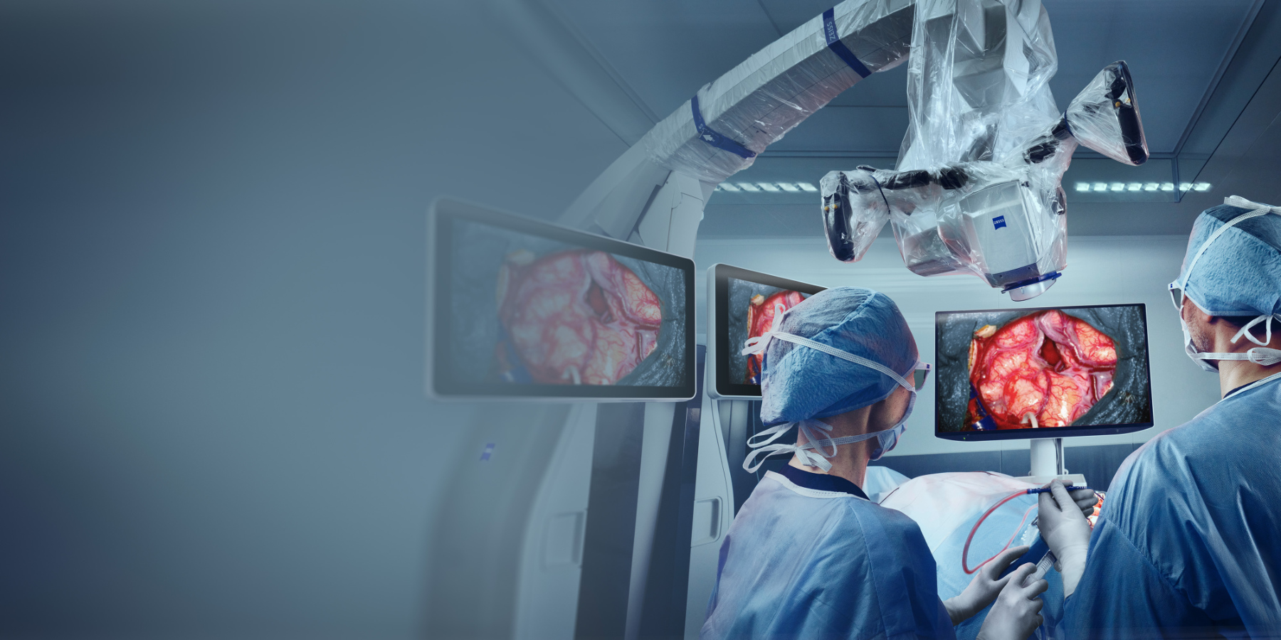
Complex surgeries in the brain and spine require the highest standards in digital and optical visualization. The new ZEISS KINEVO 900 S allows you to visualize small anatomical landmarks or tissue color differences with even more confidence thanks to best digital visualization capabilities and latest 4K 3D camera technology.
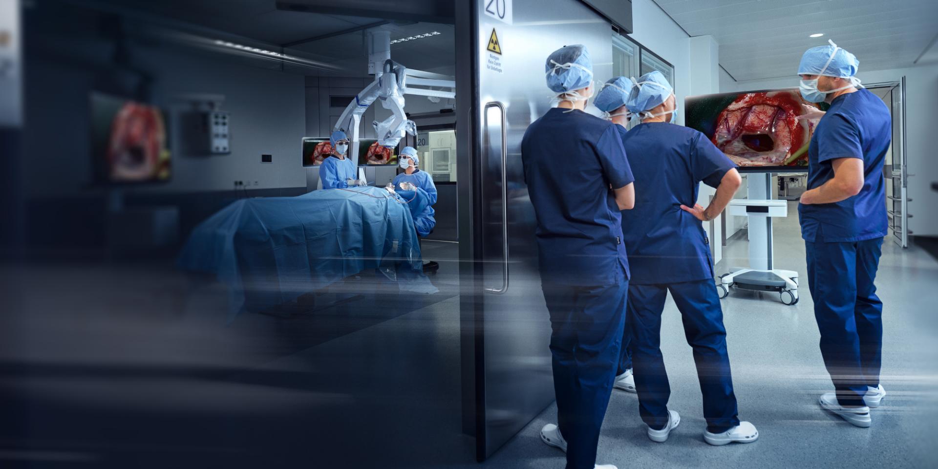
KINEVO 900 S provides a new level of visualization that allows students and assistants to precisely observe on a 4K 3D screen what the operating surgeon sees, offering superior teaching possibilities.
Experience a new dimension of clarity – ZEISS KINEVO 900 S
4K 3D technology
Work exoscopically with ease
ZEISS KINEVO 900 S offers the highest standard in digital visualization with its 4K 3D technology. By harnessing the latest technology, its enhanced 4K camera shows more visible and detailed lines, more resolution, and more colors than ever before.
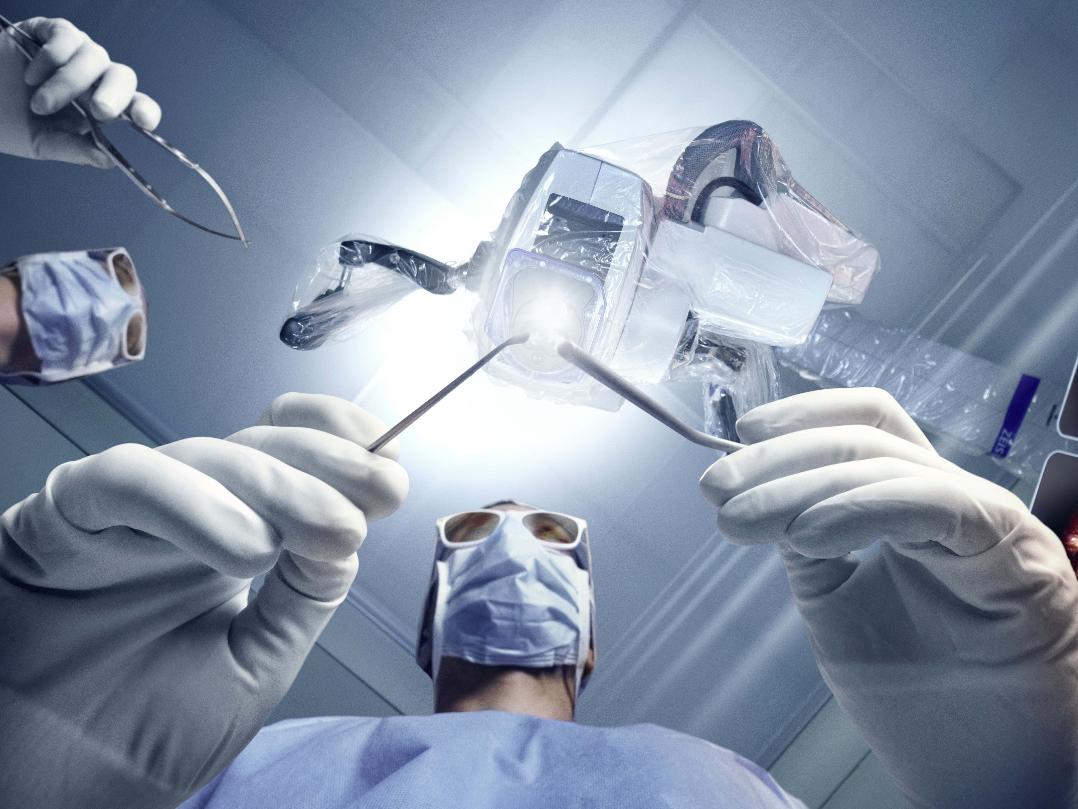
ZEISS KINEVO 900 S is not only a powerful digital solution, but it also serves as your ideal assistant for achieving an ergonomic posture during microsurgical interventions.
Our cutting-edge technology enables every team member to access the same high-resolution, three-dimensional video feed as the main surgeon. This means that students and assistants can now see what the main surgeon sees even when working through oculars and follow every step of the surgical procedure on the external monitor.
Video presets
Save valuable time during surgery
Predefined video settings on the main screen simplify your work and all other settings can be adjusted with ease.
Synchronized video settings for the camera and screen provide greater confidence and time efficiency. Working completely exoscopically is now easier than ever before.
Comparison of Enhanced Standard Mode and Brilliance Mode
What is possible with the video presets?
Adjust the slider to see the difference in the visualization during cerebellopontine angle tumor (acoustic neurinoma) surgery. While the Enhanced Standard Mode picture already delivers an optimized visualization, the Brilliance Mode adds vibrant colors enhanced contrast. This mode can be used for additional insights during the surgery and for enhanced picture quality for presentations.
ZEISS BLUE 400 S
Understand the background
The high-quality visualization of the new ZEISS BLUE 400 S, especially of the non-fluorescent parts, supports the visualization of tissue associated with Grade III & IV glioma during neurosurgeries with less frequent switching between fluorescence and white light imaging modes.
With the visualization of non-fluorescent anatomy more similar to white light impression, the understanding of anatomy on non-fluorescent tissue will increase.
Comparison of ZEISS BLUE 400 S and ZEISS BLUE 400
What is possible with ZEISS BLUE 400 S?
Adjust the slider to see the difference in the visualization during glioma surgery. The high-quality visualization of the new ZEISS BLUE 400 S, especially of the non-fluorescent parts, supports less frequent switching between fluorescence and white light imaging modes.
Which additional fluorescence options do we offer in the ZEISS KINEVO 900 S?
Experience enhanced efficiency and precision in your medical procedures with the new DepthPro mode. This solution provides greater depth of field in both the digital and optical mode, allowing you to choose the appropriate settings with more options and see everything in your field of view focused with just a click, whenever needed.
Achieve optimal visualization of tissues and structures during your procedures with our innovative solution, delivering unparalleled accuracy and precision.
Comparison of the Normal Mode and DepthPro Mode
What is possible with DepthPro?
Adjust the slider to see the difference in the visualization during cerebellopontine angle tumor (acoustic neurinoma) surgery. The DepthPro mode adds additional depth of field during this surgery to see more of the surgical field in focus, especially helpful when working in this deep corridor.
HMDmd CR3
Innovating Surgical Visualization
Experience the cutting-edge capabilities of the HMDmd CR31 wearable monitor, designed to display high-definition images in both 2D and 3D. It has a direct plug & play with the new KINEVO 900 S and improved ergonomics.
This innovative, lightweight device (250g/9oz) is specifically tailored for use in hospital environments, providing medical professionals with a portable and convenient solution for medical imaging display. Whether in operating rooms, ambulatory surgery centers or other clinical settings, the HMDmd CR3 offers versatility and mobility.
Cobotic Assistant
for a better performance with robotics
What’s a cobotic assistant and what can it do for you? Collaborative robotics, known as cobotics, fosters a working partnership between you and the ZEISS KINEVO 900 S. It offers an uninterrupted surgical workflow by automatically centering the focus area, switching between fluorescence modes when prompted, or capturing pictures and videos, helping you to keep your hands in the surgical field.
Keep your focus – ZEISS KINEVO 900 S
AutoCenter
Stay in focus
Especially during cases with high magnification needs, it might be necessary to touch the device for the numerous amounts of small positional changes during surgery, distracting your focus.
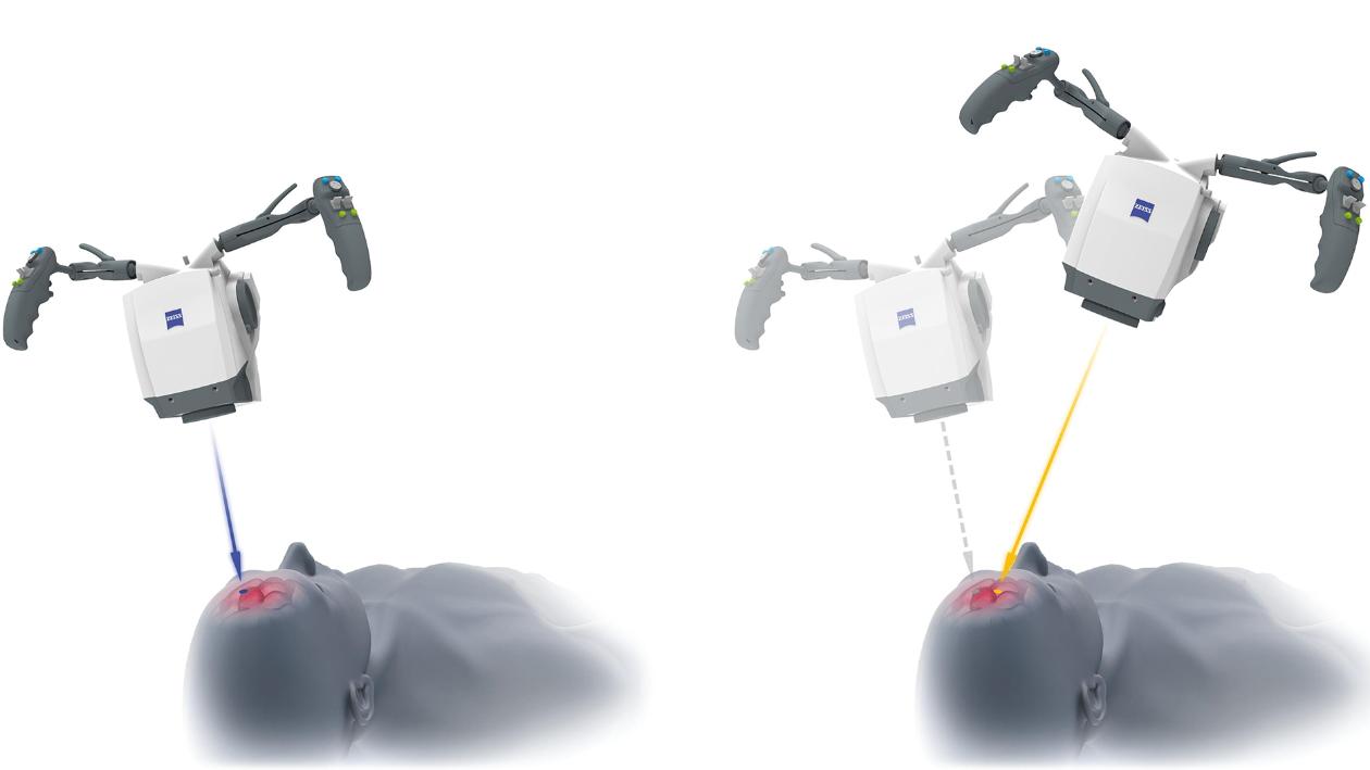
The new AutoCenter function assists you by bringing the tissue you are currently visualizing to the center of the field of view for optimized illumination and visualization, all hands-free. It identifies surgical tools within the live video and centers the field of view to the tips of the tools by using the motorized xy-functionality. This enables hands-free positioning and ensures optimized illumination, visualization, and documentation of the surgical anatomy in the field of view.
How does the AutoCenter function work?
With the touch of a button, the new AutoCenter function enables automatic re-centering of the device, providing effortless and precise adjustments. The perceived center is determined by a closed AI-trained algorithm.
As soon as an instrument is detected, you can trigger the automatic centering via the Foot Control Panel. If multiple instruments are detected, the center between their tips is automatically calculated and centered. Centering is executed by a planar xy-movement of the robotic arm of the KINEVO 900 S. This enables you to continue surgical treatment with both hands in the situs.
Voice Assistant
Say “Hey KINEVO”
Save time but still access important functions on your device. But how can you achieve both? By simply giving your ZEISS KINEVO 900 S voice commands with the ZEISS Voice Assistant – Hey KINEVO!
Experience the same convenience as with voice assistants from your everyday life – with the innovative technology by ZEISS. In keeping with the trend toward digital technology in the OR and elsewhere, a unique Voice Assistant functionality has been implemented in ZEISS KINEVO 900 S, which allows intuitive interaction with the system by using voice commands. This lets you concentrate on your surgery without having to change the device position manually or change the fluorescence mode.
What is possible with the new Voice Assistant?
“Hey KINEVO, take photo” or “Hey KINEVO, start fluorescence” – the new ZEISS Voice Assistant offers various functionalities which enable a seamless working experience where no manual changes are necessary anymore. You just need to give a command by simply talking to your device. The following functionalities will be accessible via the Voice Assistant functionalities:
Photo
Video recording
Fluorescence mode changes
Position Memory
Light Intensity
QEVO
Selection of XY mode
Improved robotics
Move it effortlessly
Motors in all axes support accurate, smooth, and effortless positioning of the microscope during use. A new motorized z-movement option (Z-Mode) makes it easy to change the working distance along the optical axis when working from a digital screen.
Known from ZEISS KINEVO 900, movement functions like PointLock and PositionMemory allow for more convenient and accurate intraoperative microscope positioning.
The inertia reduction optimizes surgical ergonomics by allowing the device to be moved effortlessly. This takes the mass of the device and any attached instruments into account, allowing for smoother and more precise movement during procedures.
The Park & Height Assistant prevents collisions with the stand or other OR equipment, ensuring the safety and efficiency of your procedures.
Together, these functions will make your work as efficient and excellent as possible.
Functionalities in detail
Z-Mode
The stand’s kinematic design allows for a new movement mode of the microscope that only moves along the optical axis of the microscope, keeping the center and the size of the visualized anatomical structures. This allows you to work hands free while freeing up space between system and surgical field.
PointLock
The visualization head can be easily repositioned in this way while keeping the point of interest focused in the center of the field of view. Changing the focal point during the Pivot movement results in a so called “keyhole” movement, which is helpful for keeping an opening of the surgical channel in the center of field of view while inspecting the anatomy behind, for example.
Position Memory
Sensors in all axes allow the position of the visualization head to be precisely detected. Any position can be stored as a bookmark and recalled. This enables you to position the microscope exactly at a specific anatomical landmark in the surgical field of view, allowing you to compare a before/after situation or to continue to perform microsurgical tasks at a specific position.
Connected Intelligence
for proactive support in surgical and non-surgical tasks
Data nowadays plays an essential role in modern surgery, including in teaching and patient management. Especially in your day-to-day routine with the Robotic Visualization System, being able to easily transfer and use the data instantly across systems is essential for smooth and efficient processes.
To meet this need, ZEISS KINEVO 900 S provides access to leading digital solutions from ZEISS to simplify data management and facilitate peer collaboration and education.
Get connected – ZEISS KINEVO 900 S
ZEISS OPTIME complete+
Data-driven proactive care
Our new data-enhanced services guarantee provide an Uptime Guarantee2 of your ZEISS systems, supporting you in delivering excellent patient care with confidence.
Utilizing system data insights together with our profound service expertise, we guide you across multiple touchpoints in your daily routines for optimal results and proactive risk mitigation.
Guaranteed uptime2 with proactive services
Empowered performance through tailored support
Enhanced productivity with digital fleet management
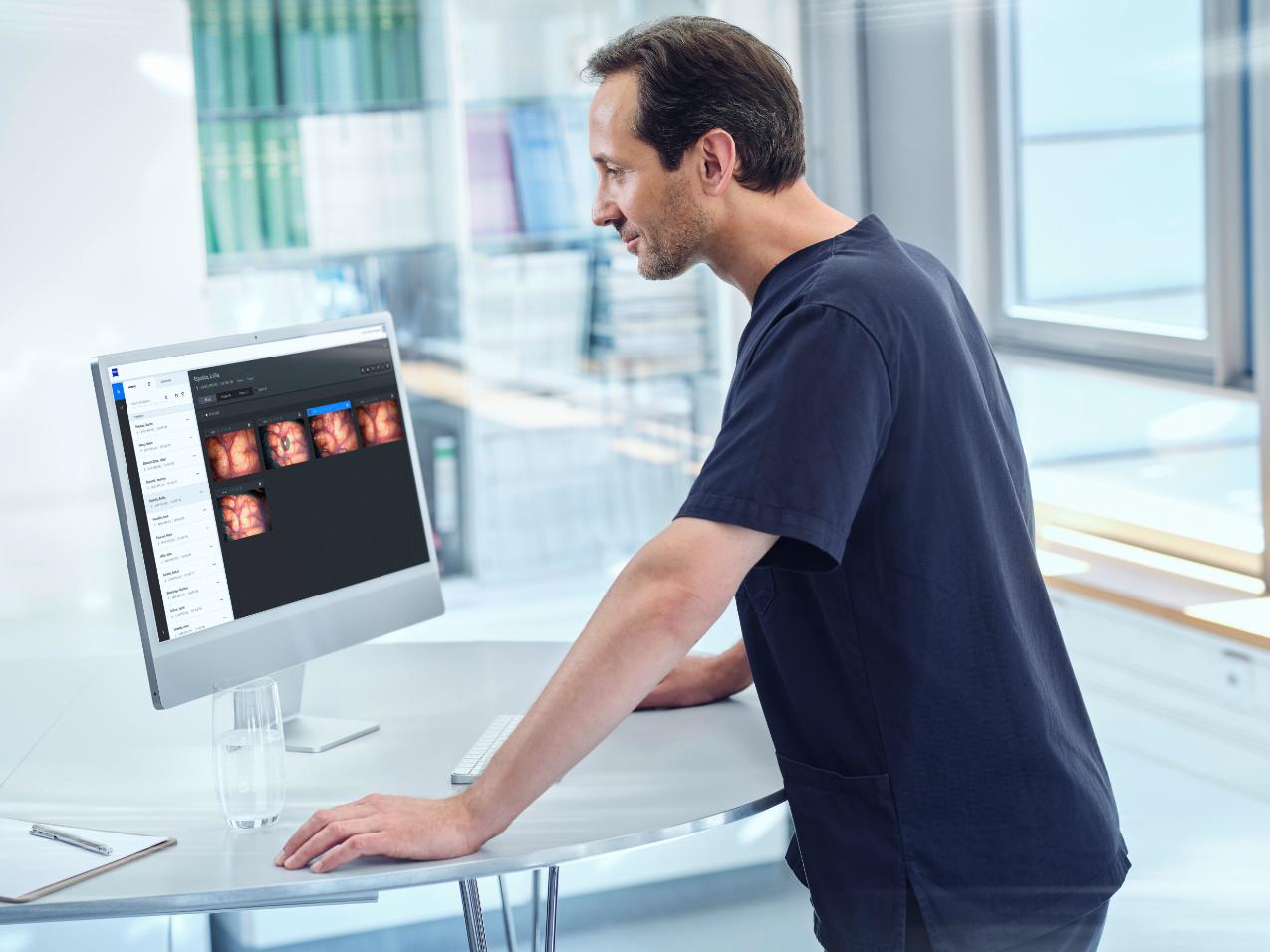
ZEISS KINEVO 900 S comes with modern software and offers integrated interfaces to all common hospital systems to be able to easily exchange data with peers. It allows you to synchronize recorded images and videos directly and securely, captured throughout surgery in a central digital location.
With this, stored data can easily be accessed, anytime, anywhere. Straightforward and remote sharing of large files and knowledge with peers will help to enhance professional collaboration and extend your network.
ZEISS Livestream
Broadcast your expertise
ZEISS Livestream offers an integrated video livestreaming solution for remote education and presentation. Simply schedule the live surgery as an upcoming webinar and send out invitation links to your remote participants to effectively stream your surgical procedure to anyone around the world in real time.
Videos
Third-party Content Blocked
The video player is blocked due to your cookie preferences. To change the settings and play the video, please click the button below and consent to use of "Functional" tracking technologies.
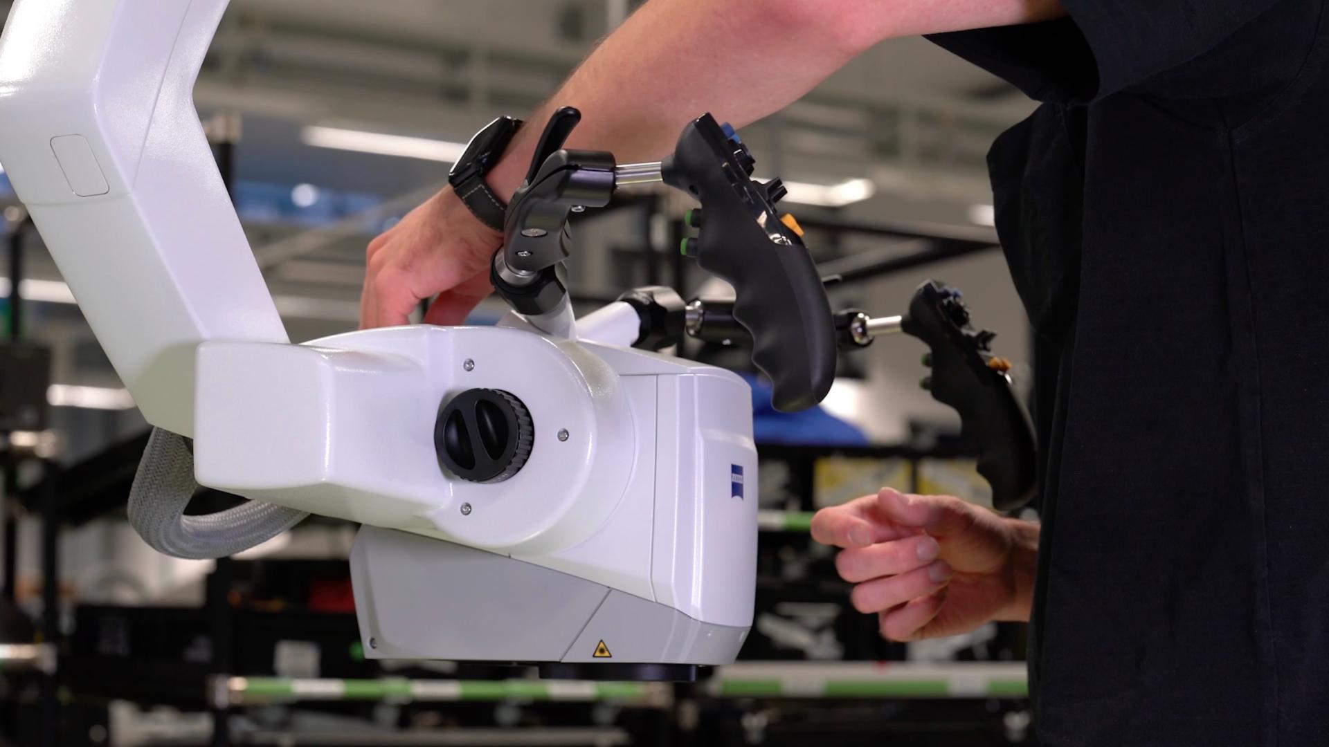
Follow the ZEISS KINEVO 900 S on its journey from the ZEISS factory to the first university hospitals
Third-party Content Blocked
The video player is blocked due to your cookie preferences. To change the settings and play the video, please click the button below and consent to use of "Functional" tracking technologies.
Experience the new possibilities for digital visualization and seamless robotic interactions
Technical Specifications
Read more about the technical specifications and the pre-defined packages of the ZEISS KINEVO 900 S


evolution")
evolution")