You are on our international English website. This site features our entire product portfolio worldwide. The products featured may not be available in the US. If you are a citizen from the US, please visit your country website for local information and contacts.
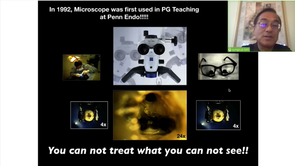
The primary pain points for dentists in removing broken endodontic instruments include limited visibility, accessibility within narrow and curved root canals and the risk of further fracturing the instrument or damaging the tooth structure.1 These challenges are critical because a retained broken instrument can lead to persistent infection, failure of the root canal treatment and potential tooth loss.2 Proper by-pass or removal is essential to prevent these complications and ensure the success of the endodontic therapy.3 High-magnification and coaxial effective illumination provided by the use of the operative microscope and specialized tools are typically required to navigate these difficulties effectively.4 The challenge of this clinical situation without microscope is shown in the picture, which demonstrate that is impossible to visualize the broken fragment without help from microscope from naked eyes perspective.
Using the microscope has revolutionized my approach to the removal of broken endodontic instruments. The enhanced magnification and illumination provide exceptional visibility, allowing for precise and effective removal, significantly reducing complications and greatly improving the success rate.
Using advanced visualization tools and specialized techniques1
Operating microscope have significantly helped tackle these challenges by providing enhanced magnification and illumination, significantly enhancing visibility and precision.4 With the help of the operating microscope, dentists can accurately detect and navigate within the narrow root canals, reducing the risk of further instrument breakage or tooth damage and increasing the success rate of instrument removal. This improved visualization given using a microscope allows for meticulous and effective instrument retrieval, by enabling dentists to see fine details that are otherwise invisible to the naked eye, thus increasing the success rate of endodontic treatments and ensuring better patient outcomes.2,3
Cheung G S. Instrument fracture: mechanisms, removal of fragments, and clinical outcomes. Endod Topics 2009;16:1–26.
2
Panitvisai P, Parunnit P, Sathorn C, Messer H H. Impact of a retained instrument on treatment outcome: a systematic and meta analysis. J Endod 2010;36:775–780.
3
Parashos, P, Messer HH. Rotary NiTi instrument fracture and its consequences. J Endod 2010;32:1031-1043.
4
Terauchi Y, Ali WT, Abielhassan MM. Present status and future directions: Removal of fractured instruments. Internat Endod J, 2022;55(Suppl. 3):685–709.