You are on our international English website. This site features our entire product portfolio worldwide. The products featured may not be available in the US. If you are a citizen from the US, please visit your country website for local information and contacts.
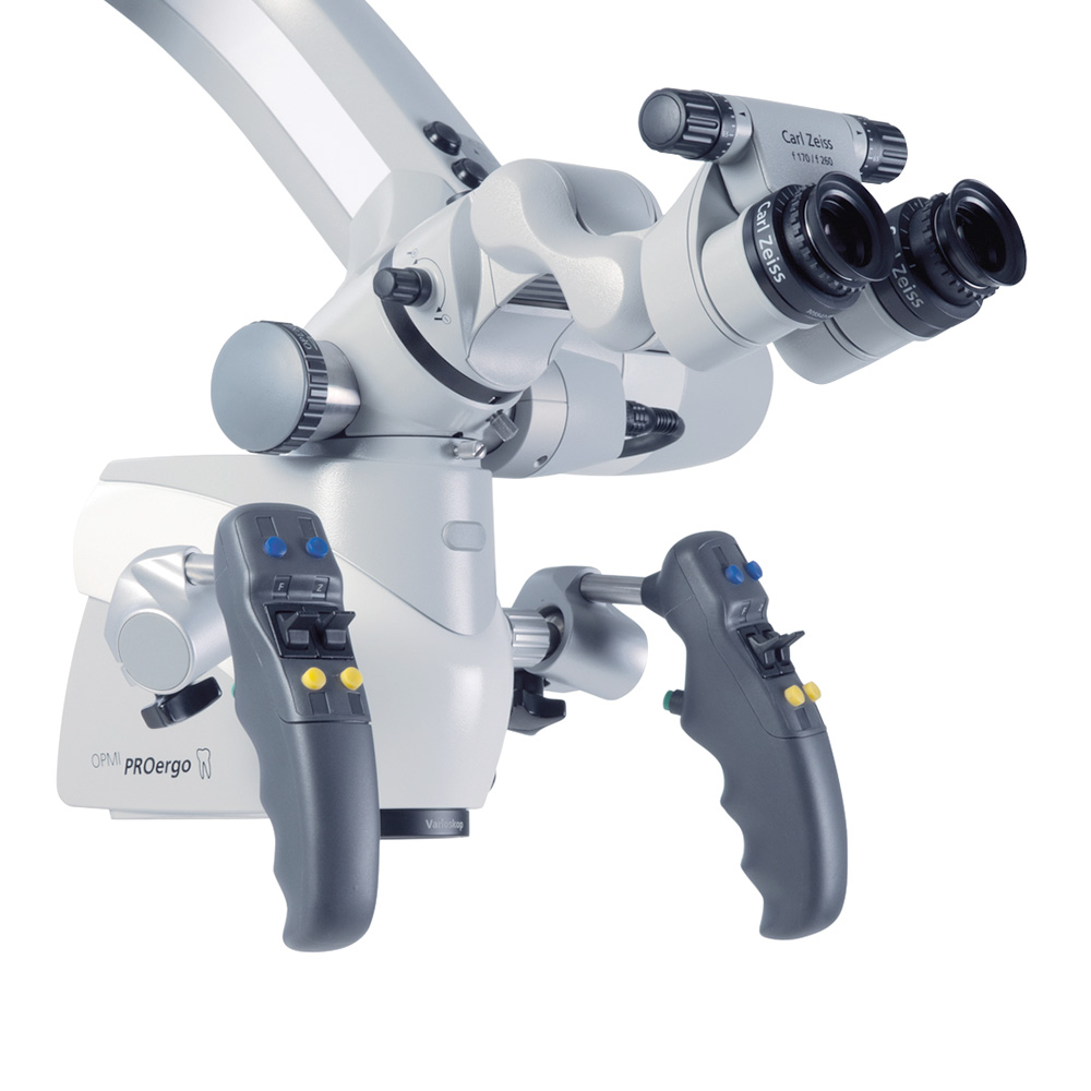
Endodontic re-treatment is considered to be one of the most challenging procedures in endodontics. In these situations, the OPMI is essential.
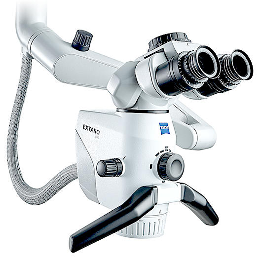
It is almost impossible to find all root canals and to see the necessary level of detail without a dental microscope. When re-treatment is required due to missed canals, broken instruments or defective fillings, the magnification and the light modes of EXTARO 300 are crucial for me to carry out the work steps precisely and ensure that the treatment is as minimally invasive as possible.
Benefits of using the OPMI on non-surgical re-treatment2
Removal of existing restorations, posts and core materials (especially useful for removal of composite cores)
Image courtesy: Dr. Tony Druttman, UK
Image courtesy: Dr. Tony Druttman, UK
Image courtesy: Dr. Tony Druttman, UK
Image courtesy: Dr. Tony Druttman, UK
Removing fractured instruments and evaluating files to avoid breakage
File evaluation
Evaluation of stainless-steel hand and NiTi rotary files under magnification and enhanced illumination is an excellent and quick way to determine if files are weakening and are at risk of separating. The dentist should look for overwound file flutes (flutes too close to each other) or unwinding flutes (the space between the flutes increases, which makes it appear shiny under enhanced lighting). Identifying this helps reduce the chance of file separation. It is much easier to identify these weak points in a file under magnification.
Image courtesy: Dr. Tony Druttman, UK
Image courtesy: Dr. Tony Druttman, UK
Image courtesy: Dr. Tony Druttman, UK
Image courtesy: Dr. Tony Druttman, UK
Evaluation and repair of perforations
Evaluation and management of perforations
The ability to visualize and determine the exact extent of a perforation helps determine treatment options and prognosis and makes it possible to repair the site.
Apical plug with MTA
MTA is an excellent material for repairing perforations and sealing large apical foramina. The material can be placed with a great deal of control when using the OPMI to ensure that there are no voids.
Image courtesy: Dr. Tony Druttman, UK
Image courtesy: Dr. Tony Druttman, UK
Image courtesy: Dr. Tony Druttman, UK
Image courtesy: Dr. Tony Druttman, UK
Image courtesy: Dr. Tony Druttman, UK
Image courtesy: Dr. Tony Druttman, UK
Removal of existing root filling materials
Image courtesy: Dr. Tony Druttman, UK
Image courtesy: Dr. Tony Druttman, UK
Image courtesy: Dr. Tony Druttman, UK
Image courtesy: Dr. Tony Druttman, UK
Image courtesy: Dr. Tony Druttman, UK
Image courtesy: Dr. Tony Druttman, UK
Image courtesy: Dr. Tony Druttman, UK
Image courtesy: Dr. Tony Druttman, UK
Removal of necrotic tissue and residual root filling materials after re-preparation of the root canals
Removal of tissue from the isthmus between the mesial canals of a lower molar using ultrasonically energised K-files.