

Examen
Visualización de cataratas densas y medición precisa


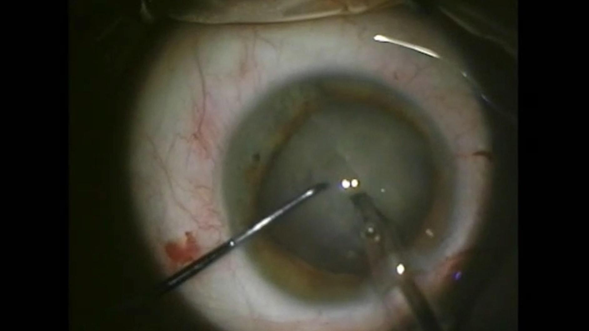
Mujer de 64 años con una catarata blanca densa intumescente
Cribado de patologías retinales en cirugía de cataratas


ZEISS IOLMaster 700
ZEISS IOLMaster 700 se ha diseñado para optimizar la eficiencia del flujo de trabajo, incluso al manipular cataratas maduras, gracias a:
- Tasa de penetración de cataratas de hasta el 99 %, que reduce la necesidad de ultrasonidos en un 92 %
- Un tiempo de medición de <45 segundos para ambos ojos7
- El examen de fijación único para identificar patologías maculares en su flujo de trabajo rutinario8


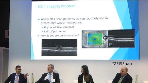
ZEISS CIRRUS 6000
ZEISS CIRRUS 6000 permite a las consultas de cataratas revelar patologías ocultas e identificar a los candidatos idóneos para la cirugía en el momento oportuno, lo cual permite una eficiencia optimizada del flujo de trabajo.
- Captura 100 000 A-scans por segundo de un campo de visión mayor de hasta 12 mm en un solo escaneo
- Los protocolos de adquisición de datos densos y los protocolos de captura oblicua aseguran la obtención de resultados diagnósticos de calidad
- Proporciona un estado objetivo de la mácula
- Ayuda a establecer las expectativas del paciente en cuanto al resultado visual de la cirugía.


-
1
S.Garg, Surgeons meet challenges of removing rock-hard cataracts, Ocular Surgery News 2018, https://www.healio.com/news/ophthalmology/20181010/surgeons-meet-challenges-of-removing-rockhard-cataracts.
-
2
U. Devgan, Dense brunescent cataracts present surgical challenges, Ocular Surgery News 2011, https://www.healio.com/news/ophthalmology/20120331/dense-brunescent-cataracts-present-surgical-challenges.
-
3
A.Brissette, OCT Is Indispensable for Pre-op Cataract Evaluation, Opthalmology Management 2019, https://www.ophthalmologymanagement.com/newsletters/insights-into-integrated-diagnostic-imaging/may-2019.
-
4
Hirnschall N et al. Macular disease detection with a swept-source optical coherence tomography-based biometry device in patients scheduled for cataract surgery. J Cataract Refract Surg. abril de 2016;42(4):530-6.
-
5
Hirnschall N et al. Enhanced Penetration for Axial Length Measurement of Eyes with Dense Cataracts Using Swept Source Optical Coherence Tomography: A Consecutive Observational Study. Ophthalmol Ther 2018;7:119-124.
-
6
R. Varsits, N. Hirnschall, B. Doeller, O. Findl; Increasing the number of successful axial eye length measurements using swept-source optical coherence tomography technology compared to conventional optical biometry; presentado en ESCSR 2016.
-
7
En función del estado del ojo y de la experiencia del cirujano.
-
8
Claramente, ZEISS IOLMaster 700 no está concebido para realizar diagnósticos. Los resultados deben ser verificados y las patologías deben diagnosticarse mediante una OCT de retina específica.
-
9
M. Colvard, Phacoemulsification of the rock hard cataract, Eyeworld 2012, https://www.eyeworld.org/article-phacoemulsification-of-the-rock-hard.
-
10
http://corporate.ewreplay.org/?v=6122663037001.
-
11
R. J. Olson, MD, Salt Lake City, Review of Ophthalmology, PUBLICADO 15 DE ENERO DE 2005 Demystifying Dysphotopsia.