Webinaire à la demande
Nouveau flux de tâches neurochirurgical pour les patients atteints de tumeurs cérébrales
7 décembre 2020
· 102 MIN VISIONNAGE
![]({"xsmall":"https://www.zeiss.com/content/dam/med-hcp/reference-master/myzeiss/peer-insights/free/pathology/509-neurosurgical-workflow-for-patients-with-brain-tumors/walavan-sivakumar.jpg/_jcr_content/renditions/original.image_file.100.100.0,10,255,265.file/walavan-sivakumar.jpg","small":"https://www.zeiss.com/content/dam/med-hcp/reference-master/myzeiss/peer-insights/free/pathology/509-neurosurgical-workflow-for-patients-with-brain-tumors/walavan-sivakumar.jpg/_jcr_content/renditions/original.image_file.255.255.0,10,255,265.file/walavan-sivakumar.jpg","medium":"https://www.zeiss.com/content/dam/med-hcp/reference-master/myzeiss/peer-insights/free/pathology/509-neurosurgical-workflow-for-patients-with-brain-tumors/walavan-sivakumar.jpg/_jcr_content/renditions/original.image_file.255.255.0,10,255,265.file/walavan-sivakumar.jpg","large":"https://www.zeiss.com/content/dam/med-hcp/reference-master/myzeiss/peer-insights/free/pathology/509-neurosurgical-workflow-for-patients-with-brain-tumors/walavan-sivakumar.jpg/_jcr_content/renditions/original.image_file.255.255.0,10,255,265.file/walavan-sivakumar.jpg","xlarge":"https://www.zeiss.com/content/dam/med-hcp/reference-master/myzeiss/peer-insights/free/pathology/509-neurosurgical-workflow-for-patients-with-brain-tumors/walavan-sivakumar.jpg/_jcr_content/renditions/original.image_file.255.255.0,10,255,265.file/walavan-sivakumar.jpg","xxlarge":"https://www.zeiss.com/content/dam/med-hcp/reference-master/myzeiss/peer-insights/free/pathology/509-neurosurgical-workflow-for-patients-with-brain-tumors/walavan-sivakumar.jpg/_jcr_content/renditions/original.image_file.255.255.0,10,255,265.file/walavan-sivakumar.jpg","max":"https://www.zeiss.com/content/dam/med-hcp/reference-master/myzeiss/peer-insights/free/pathology/509-neurosurgical-workflow-for-patients-with-brain-tumors/walavan-sivakumar.jpg/_jcr_content/renditions/original.image_file.255.255.0,10,255,265.file/walavan-sivakumar.jpg"})
Auteur
Walavan Sivakumar, MD
Neurochirurgien au Pacific Neuroscience Institute, Californie, États-Unis
![]({"xsmall":"https://www.zeiss.com/content/dam/med-hcp/reference-master/myzeiss/peer-insights/free/pathology/555-advancements-in-local-brain-tumor-treatment/nakaji.png/_jcr_content/renditions/original.image_file.100.100.0,14,261,275.file/nakaji.png","small":"https://www.zeiss.com/content/dam/med-hcp/reference-master/myzeiss/peer-insights/free/pathology/555-advancements-in-local-brain-tumor-treatment/nakaji.png/_jcr_content/renditions/original.image_file.261.261.0,14,261,275.file/nakaji.png","medium":"https://www.zeiss.com/content/dam/med-hcp/reference-master/myzeiss/peer-insights/free/pathology/555-advancements-in-local-brain-tumor-treatment/nakaji.png/_jcr_content/renditions/original.image_file.261.261.0,14,261,275.file/nakaji.png","large":"https://www.zeiss.com/content/dam/med-hcp/reference-master/myzeiss/peer-insights/free/pathology/555-advancements-in-local-brain-tumor-treatment/nakaji.png/_jcr_content/renditions/original.image_file.261.261.0,14,261,275.file/nakaji.png","xlarge":"https://www.zeiss.com/content/dam/med-hcp/reference-master/myzeiss/peer-insights/free/pathology/555-advancements-in-local-brain-tumor-treatment/nakaji.png/_jcr_content/renditions/original.image_file.261.261.0,14,261,275.file/nakaji.png","xxlarge":"https://www.zeiss.com/content/dam/med-hcp/reference-master/myzeiss/peer-insights/free/pathology/555-advancements-in-local-brain-tumor-treatment/nakaji.png/_jcr_content/renditions/original.image_file.261.261.0,14,261,275.file/nakaji.png","max":"https://www.zeiss.com/content/dam/med-hcp/reference-master/myzeiss/peer-insights/free/pathology/555-advancements-in-local-brain-tumor-treatment/nakaji.png/_jcr_content/renditions/original.image_file.261.261.0,14,261,275.file/nakaji.png"})
Auteur
Peter Nakaji, MD
Neurochirurgien au collège de médecine de l'université de l'Arizona, États-Unis
![]({"xsmall":"https://www.zeiss.com/content/dam/med-hcp/reference-master/myzeiss/peer-insights/free/pathology/509-neurosurgical-workflow-for-patients-with-brain-tumors/jennifer-eschbacher.jpg/_jcr_content/renditions/original.image_file.100.100.0,0,350,350.file/jennifer-eschbacher.jpg","small":"https://www.zeiss.com/content/dam/med-hcp/reference-master/myzeiss/peer-insights/free/pathology/509-neurosurgical-workflow-for-patients-with-brain-tumors/jennifer-eschbacher.jpg/_jcr_content/renditions/original.image_file.350.350.0,0,350,350.file/jennifer-eschbacher.jpg","medium":"https://www.zeiss.com/content/dam/med-hcp/reference-master/myzeiss/peer-insights/free/pathology/509-neurosurgical-workflow-for-patients-with-brain-tumors/jennifer-eschbacher.jpg/_jcr_content/renditions/original.image_file.350.350.0,0,350,350.file/jennifer-eschbacher.jpg","large":"https://www.zeiss.com/content/dam/med-hcp/reference-master/myzeiss/peer-insights/free/pathology/509-neurosurgical-workflow-for-patients-with-brain-tumors/jennifer-eschbacher.jpg/_jcr_content/renditions/original.image_file.350.350.0,0,350,350.file/jennifer-eschbacher.jpg","xlarge":"https://www.zeiss.com/content/dam/med-hcp/reference-master/myzeiss/peer-insights/free/pathology/509-neurosurgical-workflow-for-patients-with-brain-tumors/jennifer-eschbacher.jpg/_jcr_content/renditions/original.image_file.350.350.0,0,350,350.file/jennifer-eschbacher.jpg","xxlarge":"https://www.zeiss.com/content/dam/med-hcp/reference-master/myzeiss/peer-insights/free/pathology/509-neurosurgical-workflow-for-patients-with-brain-tumors/jennifer-eschbacher.jpg/_jcr_content/renditions/original.image_file.350.350.0,0,350,350.file/jennifer-eschbacher.jpg","max":"https://www.zeiss.com/content/dam/med-hcp/reference-master/myzeiss/peer-insights/free/pathology/509-neurosurgical-workflow-for-patients-with-brain-tumors/jennifer-eschbacher.jpg/_jcr_content/renditions/original.image_file.350.350.0,0,350,350.file/jennifer-eschbacher.jpg"})
Auteur
Jennifer Eschbacher, MD
Neuropathologue au Barrow Neurological Institute, Phoenix, Arizona, État-Unis
![]({"xsmall":"https://www.zeiss.com/content/dam/med-hcp/reference-master/myzeiss/peer-insights/free/iort/2_1607437646358/christopher-cifarelli.jpg/_jcr_content/renditions/original.image_file.100.100.0,3,255,258.file/christopher-cifarelli.jpg","small":"https://www.zeiss.com/content/dam/med-hcp/reference-master/myzeiss/peer-insights/free/iort/2_1607437646358/christopher-cifarelli.jpg/_jcr_content/renditions/original.image_file.255.255.0,3,255,258.file/christopher-cifarelli.jpg","medium":"https://www.zeiss.com/content/dam/med-hcp/reference-master/myzeiss/peer-insights/free/iort/2_1607437646358/christopher-cifarelli.jpg/_jcr_content/renditions/original.image_file.255.255.0,3,255,258.file/christopher-cifarelli.jpg","large":"https://www.zeiss.com/content/dam/med-hcp/reference-master/myzeiss/peer-insights/free/iort/2_1607437646358/christopher-cifarelli.jpg/_jcr_content/renditions/original.image_file.255.255.0,3,255,258.file/christopher-cifarelli.jpg","xlarge":"https://www.zeiss.com/content/dam/med-hcp/reference-master/myzeiss/peer-insights/free/iort/2_1607437646358/christopher-cifarelli.jpg/_jcr_content/renditions/original.image_file.255.255.0,3,255,258.file/christopher-cifarelli.jpg","xxlarge":"https://www.zeiss.com/content/dam/med-hcp/reference-master/myzeiss/peer-insights/free/iort/2_1607437646358/christopher-cifarelli.jpg/_jcr_content/renditions/original.image_file.255.255.0,3,255,258.file/christopher-cifarelli.jpg","max":"https://www.zeiss.com/content/dam/med-hcp/reference-master/myzeiss/peer-insights/free/iort/2_1607437646358/christopher-cifarelli.jpg/_jcr_content/renditions/original.image_file.255.255.0,3,255,258.file/christopher-cifarelli.jpg"})
Auteur
Christopher Cifarelli, MD, PhD
Directeur de radio-oncologie, programme de radiochirurgie par Gamma Knife de l'université de Virginie-Occidentale, États-Unis
![]({"xsmall":"https://www.zeiss.com/content/dam/med-hcp/reference-master/myzeiss/peer-insights/free/neurosurgery/1_1608037835433/klaus-henning-kahl.jpg/_jcr_content/renditions/original.image_file.100.100.0,31,255,286.file/klaus-henning-kahl.jpg","small":"https://www.zeiss.com/content/dam/med-hcp/reference-master/myzeiss/peer-insights/free/neurosurgery/1_1608037835433/klaus-henning-kahl.jpg/_jcr_content/renditions/original.image_file.255.255.0,31,255,286.file/klaus-henning-kahl.jpg","medium":"https://www.zeiss.com/content/dam/med-hcp/reference-master/myzeiss/peer-insights/free/neurosurgery/1_1608037835433/klaus-henning-kahl.jpg/_jcr_content/renditions/original.image_file.255.255.0,31,255,286.file/klaus-henning-kahl.jpg","large":"https://www.zeiss.com/content/dam/med-hcp/reference-master/myzeiss/peer-insights/free/neurosurgery/1_1608037835433/klaus-henning-kahl.jpg/_jcr_content/renditions/original.image_file.255.255.0,31,255,286.file/klaus-henning-kahl.jpg","xlarge":"https://www.zeiss.com/content/dam/med-hcp/reference-master/myzeiss/peer-insights/free/neurosurgery/1_1608037835433/klaus-henning-kahl.jpg/_jcr_content/renditions/original.image_file.255.255.0,31,255,286.file/klaus-henning-kahl.jpg","xxlarge":"https://www.zeiss.com/content/dam/med-hcp/reference-master/myzeiss/peer-insights/free/neurosurgery/1_1608037835433/klaus-henning-kahl.jpg/_jcr_content/renditions/original.image_file.255.255.0,31,255,286.file/klaus-henning-kahl.jpg","max":"https://www.zeiss.com/content/dam/med-hcp/reference-master/myzeiss/peer-insights/free/neurosurgery/1_1608037835433/klaus-henning-kahl.jpg/_jcr_content/renditions/original.image_file.255.255.0,31,255,286.file/klaus-henning-kahl.jpg"})
Auteur
Henning Kahl, MD
Radio-oncologue à l'hôpital universitaire d'Augsbourg, Allemagne
RÉSUMÉ
Aperçu d'un nouveau flux de tâches neurochirurgical pour les patients atteints de tumeurs cérébrales
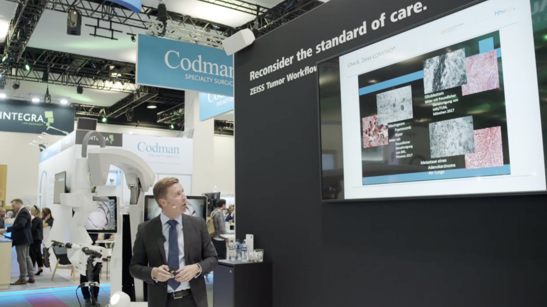
Au cours de ce webinaire multidisciplinaire ZEISS, des neurochirurgiens, des radio-oncologues et un neuropathologue partagent leurs points de vue sur l'application des technologies de ZEISS Tumor Workflow.
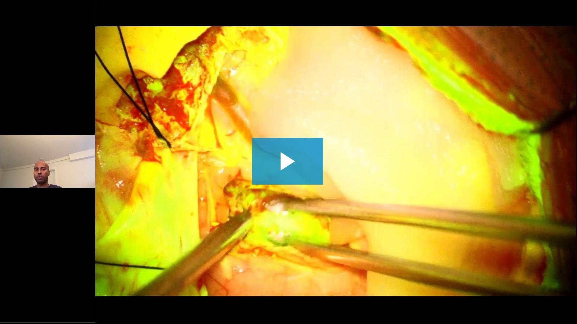
Les sujets suivants sont couverts : l'utilisation de la fluorescéine pour les tumeurs cérébrales, les expériences cliniques avec le système d'endomicroscopie confocale in vivo ZEISS CONVIVO d'un point de vue neurochirurgical (à partir de 24:05) et neuropathologique (à partir de 37:00) ainsi que le point de vue d'un radio-oncologue (à partir de 1:01:00) sur le rôle de la RTPO dans le traitement des métastases cérébrales et des glioblastomes.
![]({"xsmall":"https://www.zeiss.com/content/dam/med-hcp/reference-master/myzeiss/peer-insights/free/pathology/509-neurosurgical-workflow-for-patients-with-brain-tumors/509-neurosurgical-workflow-for-patients-with-brain-tumors_preview.jpg/_jcr_content/renditions/original.image_file.100.56.6,5,1919,1081.file/509-neurosurgical-workflow-for-patients-with-brain-tumors_preview.jpg","small":"https://www.zeiss.com/content/dam/med-hcp/reference-master/myzeiss/peer-insights/free/pathology/509-neurosurgical-workflow-for-patients-with-brain-tumors/509-neurosurgical-workflow-for-patients-with-brain-tumors_preview.jpg/_jcr_content/renditions/original.image_file.360.203.6,5,1919,1081.file/509-neurosurgical-workflow-for-patients-with-brain-tumors_preview.jpg","medium":"https://www.zeiss.com/content/dam/med-hcp/reference-master/myzeiss/peer-insights/free/pathology/509-neurosurgical-workflow-for-patients-with-brain-tumors/509-neurosurgical-workflow-for-patients-with-brain-tumors_preview.jpg/_jcr_content/renditions/original.image_file.768.432.6,5,1919,1081.file/509-neurosurgical-workflow-for-patients-with-brain-tumors_preview.jpg","large":"https://www.zeiss.com/content/dam/med-hcp/reference-master/myzeiss/peer-insights/free/pathology/509-neurosurgical-workflow-for-patients-with-brain-tumors/509-neurosurgical-workflow-for-patients-with-brain-tumors_preview.jpg/_jcr_content/renditions/original.image_file.1024.576.6,5,1919,1081.file/509-neurosurgical-workflow-for-patients-with-brain-tumors_preview.jpg","xlarge":"https://www.zeiss.com/content/dam/med-hcp/reference-master/myzeiss/peer-insights/free/pathology/509-neurosurgical-workflow-for-patients-with-brain-tumors/509-neurosurgical-workflow-for-patients-with-brain-tumors_preview.jpg/_jcr_content/renditions/original.image_file.1280.720.6,5,1919,1081.file/509-neurosurgical-workflow-for-patients-with-brain-tumors_preview.jpg","xxlarge":"https://www.zeiss.com/content/dam/med-hcp/reference-master/myzeiss/peer-insights/free/pathology/509-neurosurgical-workflow-for-patients-with-brain-tumors/509-neurosurgical-workflow-for-patients-with-brain-tumors_preview.jpg/_jcr_content/renditions/original.image_file.1440.810.6,5,1919,1081.file/509-neurosurgical-workflow-for-patients-with-brain-tumors_preview.jpg","max":"https://www.zeiss.com/content/dam/med-hcp/reference-master/myzeiss/peer-insights/free/pathology/509-neurosurgical-workflow-for-patients-with-brain-tumors/509-neurosurgical-workflow-for-patients-with-brain-tumors_preview.jpg/_jcr_content/renditions/original.image_file.1913.1076.6,5,1919,1081.file/509-neurosurgical-workflow-for-patients-with-brain-tumors_preview.jpg"})