

Évaluation
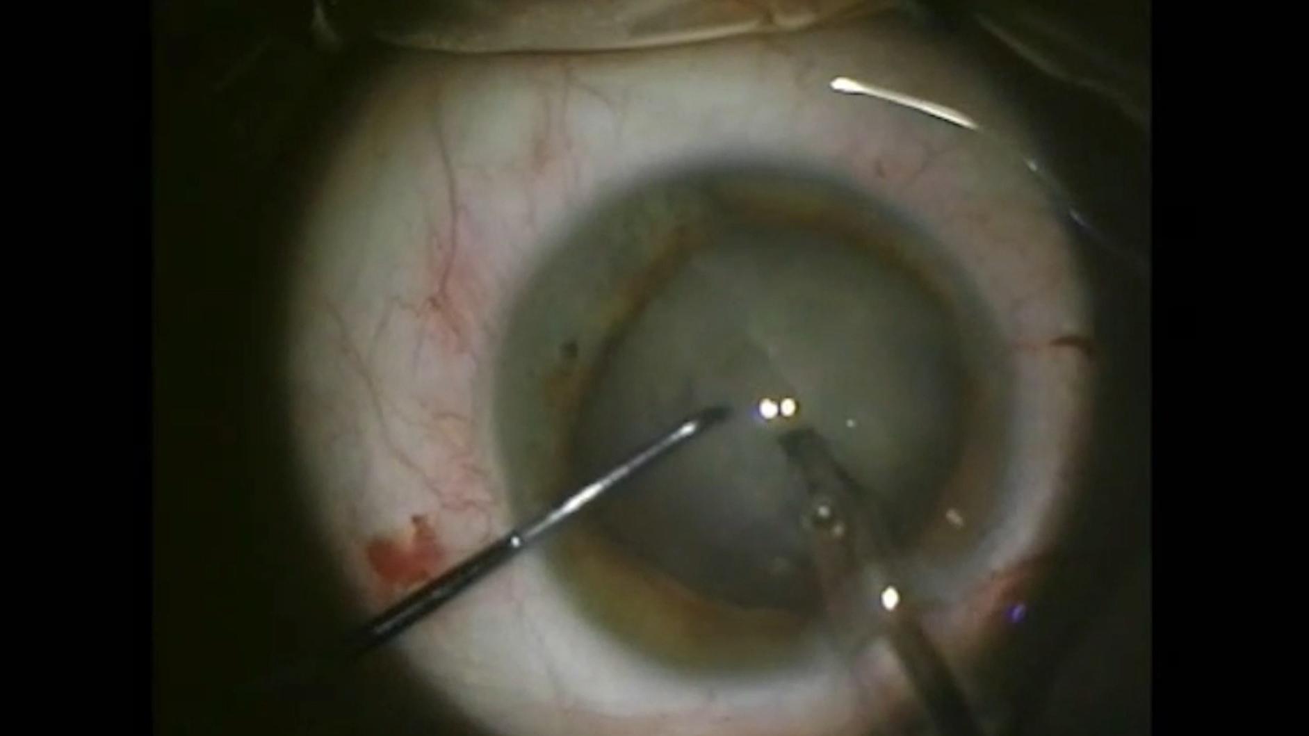
Visualisation et mesure précise des cataractes denses

Femme de 64 ans présentant une cataracte intumescente blanche et dense
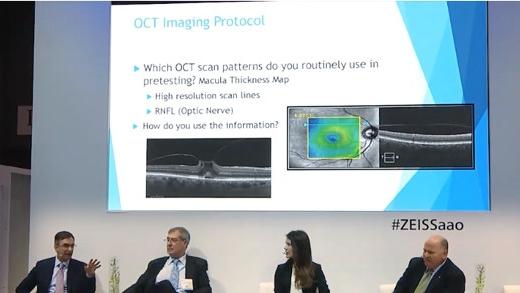
Dépistage des pathologies rétiniennes dans la chirurgie de la cataracte


ZEISS IOLMaster 700
ZEISS IOLMaster 700 est conçu pour optimiser l'efficacité du flux de tâches, même lors du traitement de cataractes denses, grâce à :
- un taux de pénétration de la cataracte pouvant atteindre 99 %, ce qui réduit de 92 % la nécessité de recourir aux ultrasons
- un temps de mesure inférieur à 45 secondes pour les deux yeux7
- la fonction Fixation Check unique pour identifier les pathologies maculaires dans votre flux de tâches habituel8


ZEISS CIRRUS 6000
ZEISS CIRRUS 6000 permet de détecter les pathologies cachées et d'identifier les candidats à une intervention chirurgicale pour un flux de tâches optimisé.
- Il effectue 100 000 A-scans par seconde pour un champ de vision plus large allant jusqu'à 12 mm, le tout en un seul passage
- Des protocoles d'acquisition de données denses et des processus de capture oblique garantissent la qualité des résultats de diagnostic
- Il permet d'évaluer de manière objective l'état de la macula
- Il aide à définir les attentes du patient quant au résultat visuel final après la chirurgie.


-
1
S.Garg, Surgeons meet challenges of removing rock-hard cataracts, Ocular Surgery News 2018, https://www.healio.com/news/ophthalmology/20181010/surgeons-meet-challenges-of-removing-rockhard-cataracts.
-
2
U. Devgan, Dense brunescent cataracts present surgical challenges, Ocular Surgery News 2011, https://www.healio.com/news/ophthalmology/20120331/dense-brunescent-cataracts-present-surgical-challenges.
-
3
A.Brissette, OCT Is Indispensable for Pre-op Cataract Evaluation, Opthalmology Management 2019, https://www.ophthalmologymanagement.com/newsletters/insights-into-integrated-diagnostic-imaging/may-2019.
-
4
Hirnschall N et al. Macular disease detection with a swept-source optical coherence tomography-based biometry device in patients scheduled for cataract surgery. J Cataract Refract Surg. 2016 Avr;42(4):530-6.
-
5
Hirnschall N et al. Enhanced Penetration for Axial Length Measurement of Eyes with Dense Cataracts Using Swept Source Optical Coherence Tomography: A Consecutive Observational Study. Ophthalmol Ther 2018;7:119-124.
-
6
R. Varsits, N. Hirnschall, B. Doeller, O. Findl; Increasing the number of successful axial eye length measurements using swept-source optical coherence tomography technology compared to conventional optical biometry; présentée à l'ESCSR 2016.
-
7
Selon l'état des yeux et l'expérience de l'opérateur.
-
8
ZEISS IOLMaster 700 n'est clairement pas prévu pour une utilisation diagnostique. Les résultats doivent être vérifiés et les pathologies diagnostiquées avec un OCT de la rétine dédié.
-
9
M. Colvard, Phacoemulsification of the rock hard cataract, Eyeworld 2012, https://www.eyeworld.org/article-phacoemulsification-of-the-rock-hard.
-
10
http://corporate.ewreplay.org/?v=6122663037001.
-
11
Dr R. J. Olson, Salt Lake City, Review of Ophthalmology, PUBLIÉ LE 15 JANVIER 2005 Demystifying Dysphotopsia.