Webinar on demand
Nuovo flusso di lavoro neurochirurgico per pazienti con tumori cerebrali
7 dicembre 2020
· 102 MIN VISIONE
![]({"xsmall":"https://www.zeiss.com/content/dam/med-hcp/reference-master/myzeiss/peer-insights/free/pathology/509-neurosurgical-workflow-for-patients-with-brain-tumors/walavan-sivakumar.jpg/_jcr_content/renditions/original.image_file.100.100.0,10,255,265.file/walavan-sivakumar.jpg","small":"https://www.zeiss.com/content/dam/med-hcp/reference-master/myzeiss/peer-insights/free/pathology/509-neurosurgical-workflow-for-patients-with-brain-tumors/walavan-sivakumar.jpg/_jcr_content/renditions/original.image_file.255.255.0,10,255,265.file/walavan-sivakumar.jpg","medium":"https://www.zeiss.com/content/dam/med-hcp/reference-master/myzeiss/peer-insights/free/pathology/509-neurosurgical-workflow-for-patients-with-brain-tumors/walavan-sivakumar.jpg/_jcr_content/renditions/original.image_file.255.255.0,10,255,265.file/walavan-sivakumar.jpg","large":"https://www.zeiss.com/content/dam/med-hcp/reference-master/myzeiss/peer-insights/free/pathology/509-neurosurgical-workflow-for-patients-with-brain-tumors/walavan-sivakumar.jpg/_jcr_content/renditions/original.image_file.255.255.0,10,255,265.file/walavan-sivakumar.jpg","xlarge":"https://www.zeiss.com/content/dam/med-hcp/reference-master/myzeiss/peer-insights/free/pathology/509-neurosurgical-workflow-for-patients-with-brain-tumors/walavan-sivakumar.jpg/_jcr_content/renditions/original.image_file.255.255.0,10,255,265.file/walavan-sivakumar.jpg","xxlarge":"https://www.zeiss.com/content/dam/med-hcp/reference-master/myzeiss/peer-insights/free/pathology/509-neurosurgical-workflow-for-patients-with-brain-tumors/walavan-sivakumar.jpg/_jcr_content/renditions/original.image_file.255.255.0,10,255,265.file/walavan-sivakumar.jpg","max":"https://www.zeiss.com/content/dam/med-hcp/reference-master/myzeiss/peer-insights/free/pathology/509-neurosurgical-workflow-for-patients-with-brain-tumors/walavan-sivakumar.jpg/_jcr_content/renditions/original.image_file.255.255.0,10,255,265.file/walavan-sivakumar.jpg"})
Autore
Walavan Sivakumar, MD
Neurochirurgo, Pacific Neuroscience Institute, California, Stati Uniti
![]({"xsmall":"https://www.zeiss.com/content/dam/med-hcp/reference-master/myzeiss/peer-insights/free/pathology/555-advancements-in-local-brain-tumor-treatment/nakaji.png/_jcr_content/renditions/original.image_file.100.100.0,14,261,275.file/nakaji.png","small":"https://www.zeiss.com/content/dam/med-hcp/reference-master/myzeiss/peer-insights/free/pathology/555-advancements-in-local-brain-tumor-treatment/nakaji.png/_jcr_content/renditions/original.image_file.261.261.0,14,261,275.file/nakaji.png","medium":"https://www.zeiss.com/content/dam/med-hcp/reference-master/myzeiss/peer-insights/free/pathology/555-advancements-in-local-brain-tumor-treatment/nakaji.png/_jcr_content/renditions/original.image_file.261.261.0,14,261,275.file/nakaji.png","large":"https://www.zeiss.com/content/dam/med-hcp/reference-master/myzeiss/peer-insights/free/pathology/555-advancements-in-local-brain-tumor-treatment/nakaji.png/_jcr_content/renditions/original.image_file.261.261.0,14,261,275.file/nakaji.png","xlarge":"https://www.zeiss.com/content/dam/med-hcp/reference-master/myzeiss/peer-insights/free/pathology/555-advancements-in-local-brain-tumor-treatment/nakaji.png/_jcr_content/renditions/original.image_file.261.261.0,14,261,275.file/nakaji.png","xxlarge":"https://www.zeiss.com/content/dam/med-hcp/reference-master/myzeiss/peer-insights/free/pathology/555-advancements-in-local-brain-tumor-treatment/nakaji.png/_jcr_content/renditions/original.image_file.261.261.0,14,261,275.file/nakaji.png","max":"https://www.zeiss.com/content/dam/med-hcp/reference-master/myzeiss/peer-insights/free/pathology/555-advancements-in-local-brain-tumor-treatment/nakaji.png/_jcr_content/renditions/original.image_file.261.261.0,14,261,275.file/nakaji.png"})
Autore
Peter Nakaji, MD
Neurochirurgo University of Arizona College of Medicine, Stati Uniti
![]({"xsmall":"https://www.zeiss.com/content/dam/med-hcp/reference-master/myzeiss/peer-insights/free/pathology/509-neurosurgical-workflow-for-patients-with-brain-tumors/jennifer-eschbacher.jpg/_jcr_content/renditions/original.image_file.100.100.0,0,350,350.file/jennifer-eschbacher.jpg","small":"https://www.zeiss.com/content/dam/med-hcp/reference-master/myzeiss/peer-insights/free/pathology/509-neurosurgical-workflow-for-patients-with-brain-tumors/jennifer-eschbacher.jpg/_jcr_content/renditions/original.image_file.350.350.0,0,350,350.file/jennifer-eschbacher.jpg","medium":"https://www.zeiss.com/content/dam/med-hcp/reference-master/myzeiss/peer-insights/free/pathology/509-neurosurgical-workflow-for-patients-with-brain-tumors/jennifer-eschbacher.jpg/_jcr_content/renditions/original.image_file.350.350.0,0,350,350.file/jennifer-eschbacher.jpg","large":"https://www.zeiss.com/content/dam/med-hcp/reference-master/myzeiss/peer-insights/free/pathology/509-neurosurgical-workflow-for-patients-with-brain-tumors/jennifer-eschbacher.jpg/_jcr_content/renditions/original.image_file.350.350.0,0,350,350.file/jennifer-eschbacher.jpg","xlarge":"https://www.zeiss.com/content/dam/med-hcp/reference-master/myzeiss/peer-insights/free/pathology/509-neurosurgical-workflow-for-patients-with-brain-tumors/jennifer-eschbacher.jpg/_jcr_content/renditions/original.image_file.350.350.0,0,350,350.file/jennifer-eschbacher.jpg","xxlarge":"https://www.zeiss.com/content/dam/med-hcp/reference-master/myzeiss/peer-insights/free/pathology/509-neurosurgical-workflow-for-patients-with-brain-tumors/jennifer-eschbacher.jpg/_jcr_content/renditions/original.image_file.350.350.0,0,350,350.file/jennifer-eschbacher.jpg","max":"https://www.zeiss.com/content/dam/med-hcp/reference-master/myzeiss/peer-insights/free/pathology/509-neurosurgical-workflow-for-patients-with-brain-tumors/jennifer-eschbacher.jpg/_jcr_content/renditions/original.image_file.350.350.0,0,350,350.file/jennifer-eschbacher.jpg"})
Autore
Jennifer Eschbacher, MD
Neuropatologa Barrow Neurological Institute Phoenix, Arizona, Stati Uniti
![]({"xsmall":"https://www.zeiss.com/content/dam/med-hcp/reference-master/myzeiss/peer-insights/free/iort/2_1607437646358/christopher-cifarelli.jpg/_jcr_content/renditions/original.image_file.100.100.0,3,255,258.file/christopher-cifarelli.jpg","small":"https://www.zeiss.com/content/dam/med-hcp/reference-master/myzeiss/peer-insights/free/iort/2_1607437646358/christopher-cifarelli.jpg/_jcr_content/renditions/original.image_file.255.255.0,3,255,258.file/christopher-cifarelli.jpg","medium":"https://www.zeiss.com/content/dam/med-hcp/reference-master/myzeiss/peer-insights/free/iort/2_1607437646358/christopher-cifarelli.jpg/_jcr_content/renditions/original.image_file.255.255.0,3,255,258.file/christopher-cifarelli.jpg","large":"https://www.zeiss.com/content/dam/med-hcp/reference-master/myzeiss/peer-insights/free/iort/2_1607437646358/christopher-cifarelli.jpg/_jcr_content/renditions/original.image_file.255.255.0,3,255,258.file/christopher-cifarelli.jpg","xlarge":"https://www.zeiss.com/content/dam/med-hcp/reference-master/myzeiss/peer-insights/free/iort/2_1607437646358/christopher-cifarelli.jpg/_jcr_content/renditions/original.image_file.255.255.0,3,255,258.file/christopher-cifarelli.jpg","xxlarge":"https://www.zeiss.com/content/dam/med-hcp/reference-master/myzeiss/peer-insights/free/iort/2_1607437646358/christopher-cifarelli.jpg/_jcr_content/renditions/original.image_file.255.255.0,3,255,258.file/christopher-cifarelli.jpg","max":"https://www.zeiss.com/content/dam/med-hcp/reference-master/myzeiss/peer-insights/free/iort/2_1607437646358/christopher-cifarelli.jpg/_jcr_content/renditions/original.image_file.255.255.0,3,255,258.file/christopher-cifarelli.jpg"})
Autore
Christopher Cifarelli, MD, PhD
Direttore Radioterapia oncologica, West Virginia University, Gamma Knife Radiosurgery Program, Stati Uniti
![]({"xsmall":"https://www.zeiss.com/content/dam/med-hcp/reference-master/myzeiss/peer-insights/free/neurosurgery/1_1608037835433/klaus-henning-kahl.jpg/_jcr_content/renditions/original.image_file.100.100.0,31,255,286.file/klaus-henning-kahl.jpg","small":"https://www.zeiss.com/content/dam/med-hcp/reference-master/myzeiss/peer-insights/free/neurosurgery/1_1608037835433/klaus-henning-kahl.jpg/_jcr_content/renditions/original.image_file.255.255.0,31,255,286.file/klaus-henning-kahl.jpg","medium":"https://www.zeiss.com/content/dam/med-hcp/reference-master/myzeiss/peer-insights/free/neurosurgery/1_1608037835433/klaus-henning-kahl.jpg/_jcr_content/renditions/original.image_file.255.255.0,31,255,286.file/klaus-henning-kahl.jpg","large":"https://www.zeiss.com/content/dam/med-hcp/reference-master/myzeiss/peer-insights/free/neurosurgery/1_1608037835433/klaus-henning-kahl.jpg/_jcr_content/renditions/original.image_file.255.255.0,31,255,286.file/klaus-henning-kahl.jpg","xlarge":"https://www.zeiss.com/content/dam/med-hcp/reference-master/myzeiss/peer-insights/free/neurosurgery/1_1608037835433/klaus-henning-kahl.jpg/_jcr_content/renditions/original.image_file.255.255.0,31,255,286.file/klaus-henning-kahl.jpg","xxlarge":"https://www.zeiss.com/content/dam/med-hcp/reference-master/myzeiss/peer-insights/free/neurosurgery/1_1608037835433/klaus-henning-kahl.jpg/_jcr_content/renditions/original.image_file.255.255.0,31,255,286.file/klaus-henning-kahl.jpg","max":"https://www.zeiss.com/content/dam/med-hcp/reference-master/myzeiss/peer-insights/free/neurosurgery/1_1608037835433/klaus-henning-kahl.jpg/_jcr_content/renditions/original.image_file.255.255.0,31,255,286.file/klaus-henning-kahl.jpg"})
Autore
Henning Kahl, MD
Radioterapista oncologo, Ospedale Universitario di Augusta, Germania
RIEPILOGO
Approfondimenti per un nuovo flusso di lavoro neurochirurgico per pazienti con tumori cerebrali
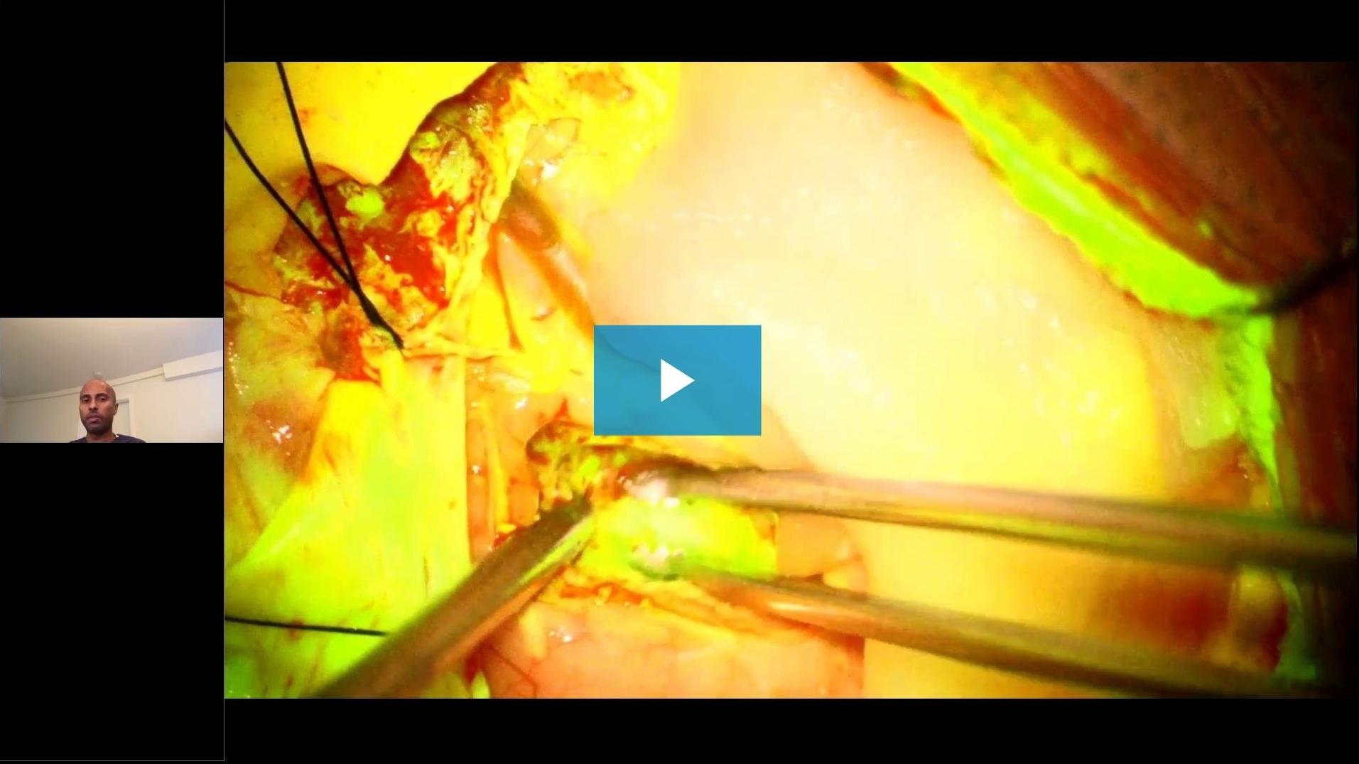
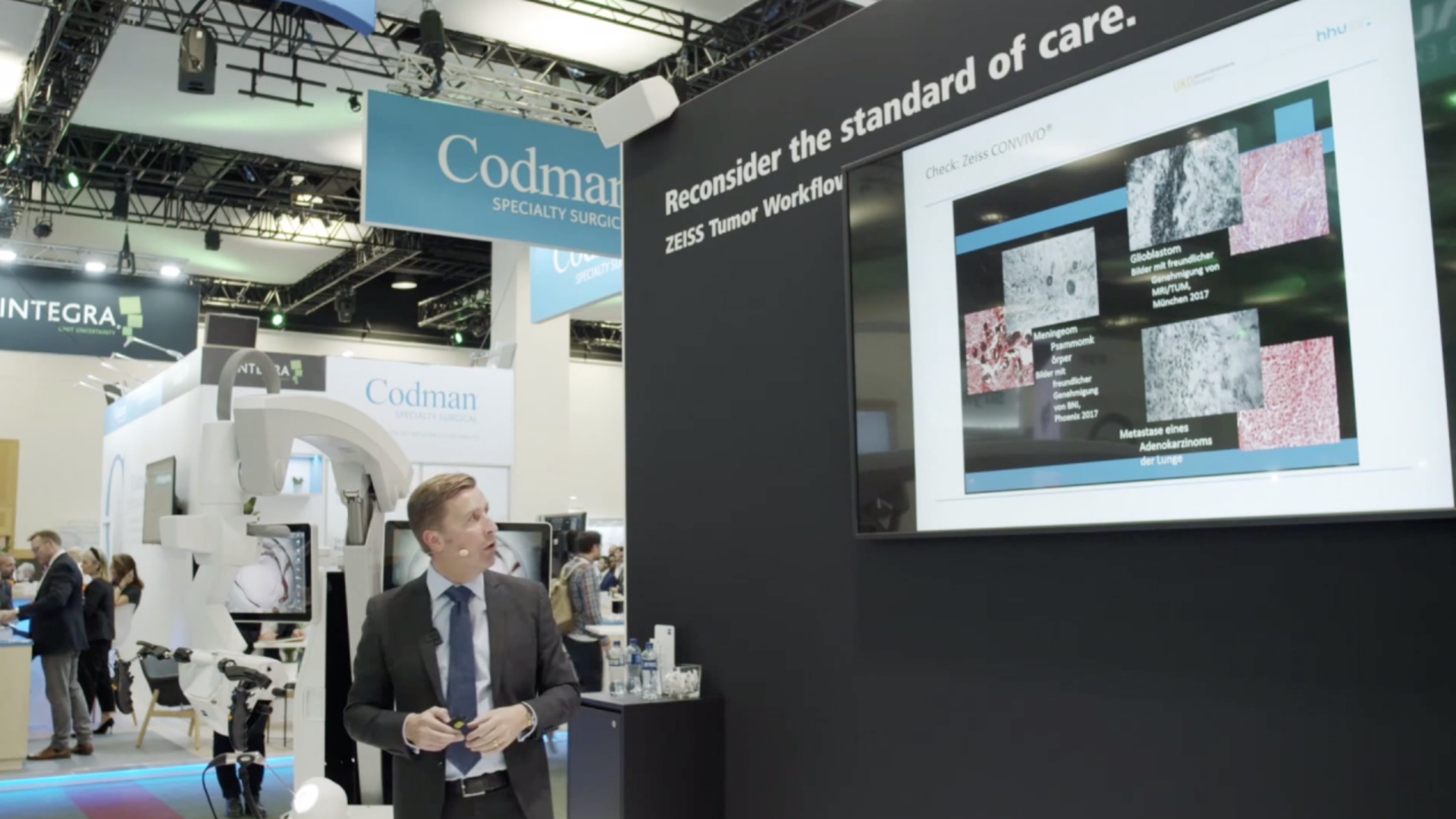
Durante questo webinar multidisciplinare ZEISS, neurochirurghi, radioterapisti oncologi e un neuropatologo condividono le loro conoscenze sull’applicazione delle tecnologie ZEISS Tumor Workflow.
Gli argomenti trattati sono: L’utilizzo della fluoresceina per i tumori cerebrali, le esperienze cliniche con il sistema di endomicroscopia confocale in vivo ZEISS CONVIVO dal punto di vista neurochirurgico (a partire dal minuto 24:05) e neuropatologico (a partire dal minuto 37:00), nonché il punto di vista degli radioterapisti oncologi (a partire dal minuto 1:01:00) sul ruolo della IORT nel trattamento delle metastasi cerebrali e del glioblastoma.
![]({"xsmall":"https://www.zeiss.com/content/dam/med-hcp/reference-master/myzeiss/peer-insights/free/pathology/509-neurosurgical-workflow-for-patients-with-brain-tumors/509-neurosurgical-workflow-for-patients-with-brain-tumors_preview.jpg/_jcr_content/renditions/original.image_file.100.56.6,5,1919,1081.file/509-neurosurgical-workflow-for-patients-with-brain-tumors_preview.jpg","small":"https://www.zeiss.com/content/dam/med-hcp/reference-master/myzeiss/peer-insights/free/pathology/509-neurosurgical-workflow-for-patients-with-brain-tumors/509-neurosurgical-workflow-for-patients-with-brain-tumors_preview.jpg/_jcr_content/renditions/original.image_file.360.203.6,5,1919,1081.file/509-neurosurgical-workflow-for-patients-with-brain-tumors_preview.jpg","medium":"https://www.zeiss.com/content/dam/med-hcp/reference-master/myzeiss/peer-insights/free/pathology/509-neurosurgical-workflow-for-patients-with-brain-tumors/509-neurosurgical-workflow-for-patients-with-brain-tumors_preview.jpg/_jcr_content/renditions/original.image_file.768.432.6,5,1919,1081.file/509-neurosurgical-workflow-for-patients-with-brain-tumors_preview.jpg","large":"https://www.zeiss.com/content/dam/med-hcp/reference-master/myzeiss/peer-insights/free/pathology/509-neurosurgical-workflow-for-patients-with-brain-tumors/509-neurosurgical-workflow-for-patients-with-brain-tumors_preview.jpg/_jcr_content/renditions/original.image_file.1024.576.6,5,1919,1081.file/509-neurosurgical-workflow-for-patients-with-brain-tumors_preview.jpg","xlarge":"https://www.zeiss.com/content/dam/med-hcp/reference-master/myzeiss/peer-insights/free/pathology/509-neurosurgical-workflow-for-patients-with-brain-tumors/509-neurosurgical-workflow-for-patients-with-brain-tumors_preview.jpg/_jcr_content/renditions/original.image_file.1280.720.6,5,1919,1081.file/509-neurosurgical-workflow-for-patients-with-brain-tumors_preview.jpg","xxlarge":"https://www.zeiss.com/content/dam/med-hcp/reference-master/myzeiss/peer-insights/free/pathology/509-neurosurgical-workflow-for-patients-with-brain-tumors/509-neurosurgical-workflow-for-patients-with-brain-tumors_preview.jpg/_jcr_content/renditions/original.image_file.1440.810.6,5,1919,1081.file/509-neurosurgical-workflow-for-patients-with-brain-tumors_preview.jpg","max":"https://www.zeiss.com/content/dam/med-hcp/reference-master/myzeiss/peer-insights/free/pathology/509-neurosurgical-workflow-for-patients-with-brain-tumors/509-neurosurgical-workflow-for-patients-with-brain-tumors_preview.jpg/_jcr_content/renditions/original.image_file.1913.1076.6,5,1919,1081.file/509-neurosurgical-workflow-for-patients-with-brain-tumors_preview.jpg"})