Presentazione scientifica
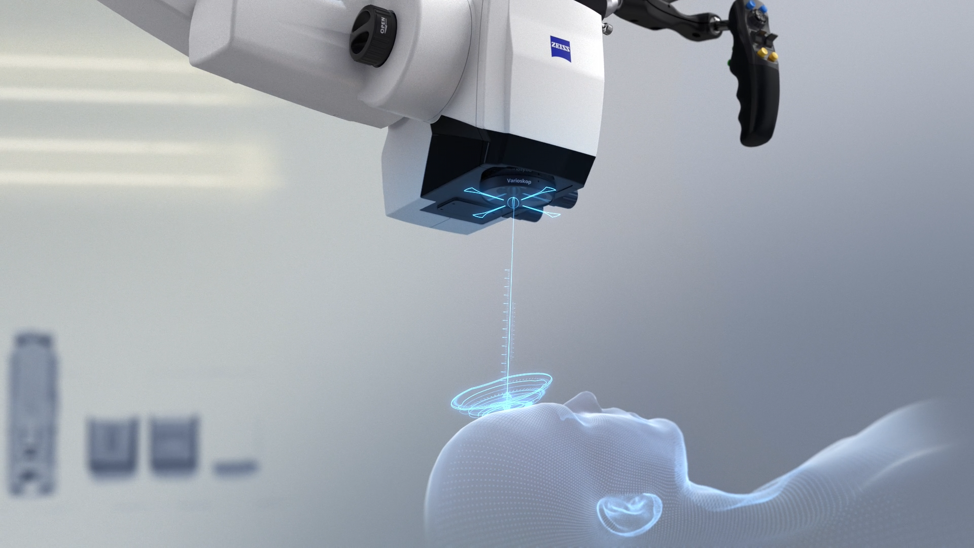
La visualizzazione avanzata incontra la radioterapia intraoperatoria
Intervento registrato in occasione dell’EANS 2019 che parla di come le tecnologie più innovative vadano a supporto del trattamento dei tumori cerebrali.
25 settembre 2019
· 23 MIN VISIONE
![]({"xsmall":"https://www.zeiss.com/content/dam/med-hcp/reference-master/myzeiss/peer-insights/free/pathology/187--combining-advanced-visualization-and-intraoperative-radiotherapy/haenggi_daniel_neu.jpg/_jcr_content/renditions/original.image_file.100.100.0,42,2268,2310.file/haenggi_daniel_neu.jpg","small":"https://www.zeiss.com/content/dam/med-hcp/reference-master/myzeiss/peer-insights/free/pathology/187--combining-advanced-visualization-and-intraoperative-radiotherapy/haenggi_daniel_neu.jpg/_jcr_content/renditions/original.image_file.360.360.0,42,2268,2310.file/haenggi_daniel_neu.jpg","medium":"https://www.zeiss.com/content/dam/med-hcp/reference-master/myzeiss/peer-insights/free/pathology/187--combining-advanced-visualization-and-intraoperative-radiotherapy/haenggi_daniel_neu.jpg/_jcr_content/renditions/original.image_file.768.768.0,42,2268,2310.file/haenggi_daniel_neu.jpg","large":"https://www.zeiss.com/content/dam/med-hcp/reference-master/myzeiss/peer-insights/free/pathology/187--combining-advanced-visualization-and-intraoperative-radiotherapy/haenggi_daniel_neu.jpg/_jcr_content/renditions/original.image_file.1024.1024.0,42,2268,2310.file/haenggi_daniel_neu.jpg","xlarge":"https://www.zeiss.com/content/dam/med-hcp/reference-master/myzeiss/peer-insights/free/pathology/187--combining-advanced-visualization-and-intraoperative-radiotherapy/haenggi_daniel_neu.jpg/_jcr_content/renditions/original.image_file.1280.1280.0,42,2268,2310.file/haenggi_daniel_neu.jpg","xxlarge":"https://www.zeiss.com/content/dam/med-hcp/reference-master/myzeiss/peer-insights/free/pathology/187--combining-advanced-visualization-and-intraoperative-radiotherapy/haenggi_daniel_neu.jpg/_jcr_content/renditions/original.image_file.1440.1440.0,42,2268,2310.file/haenggi_daniel_neu.jpg","max":"https://www.zeiss.com/content/dam/med-hcp/reference-master/myzeiss/peer-insights/free/pathology/187--combining-advanced-visualization-and-intraoperative-radiotherapy/haenggi_daniel_neu.jpg/_jcr_content/renditions/original.image_file.1920.1920.0,42,2268,2310.file/haenggi_daniel_neu.jpg"})
Autore
Univ.-Prof. Dr. med. Daniel Hänggi,
Ospedale Universitario, Düsseldorf, Germania
RIEPILOGO
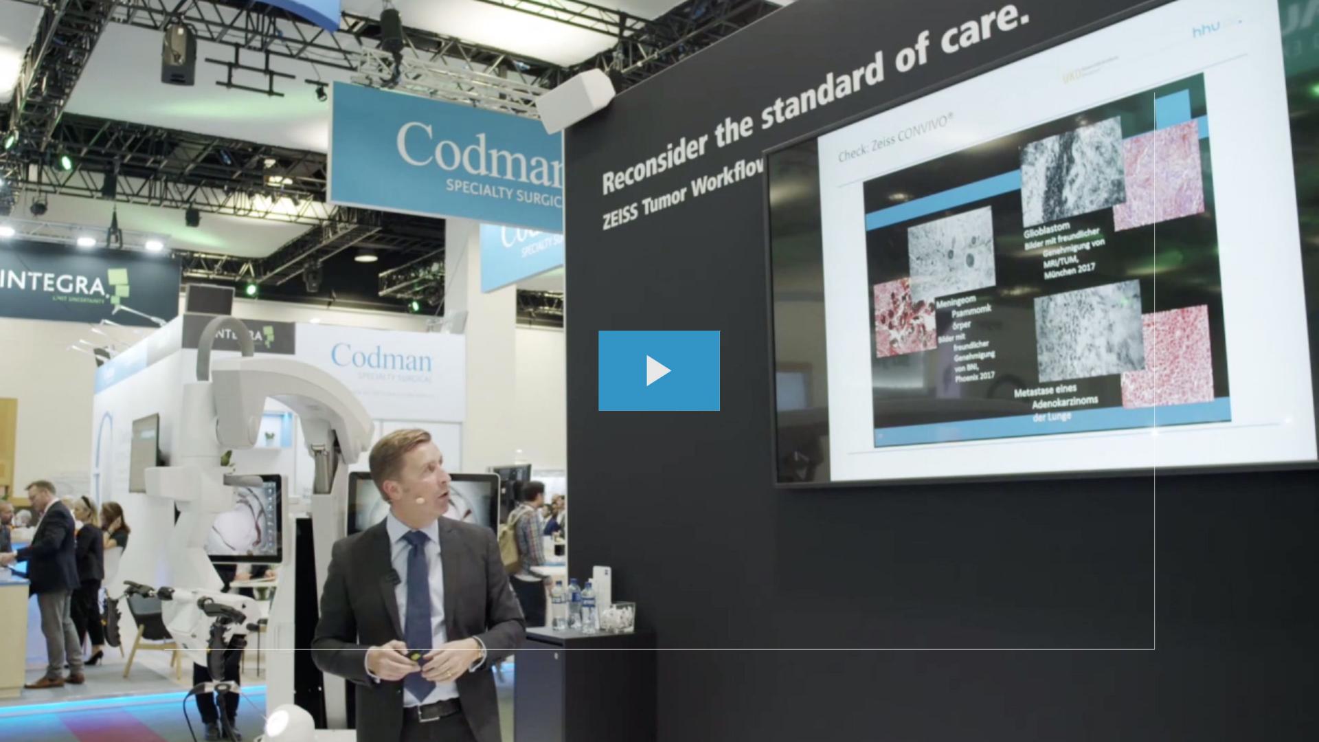
Sessione registrata di incontro con gli esperti all’EANS 2019
Ogni giorno i neurochirurghi si muovono su una linea sottile tra il raggiungimento della massima resezione del tumore e la conservazione del tessuto eloquente. In questo intervento il Dr. Hänggi condivide le sue esperienze dirette, spiegando come le ultime tecnologie supportino lui e il suo team nel trattamento dei tumori:
Visualizzazione. Controllo. Trattamento. – ZEISS Tumor Workflow.
![]({"xsmall":"https://www.zeiss.com/content/dam/med-hcp/reference-master/myzeiss/peer-insights/free/pathology/187--combining-advanced-visualization-and-intraoperative-radiotherapy/haenggi_video.jpg/_jcr_content/renditions/original.image_file.100.56.0,2,2556,1440.file/haenggi_video.jpg","small":"https://www.zeiss.com/content/dam/med-hcp/reference-master/myzeiss/peer-insights/free/pathology/187--combining-advanced-visualization-and-intraoperative-radiotherapy/haenggi_video.jpg/_jcr_content/renditions/original.image_file.360.203.0,2,2556,1440.file/haenggi_video.jpg","medium":"https://www.zeiss.com/content/dam/med-hcp/reference-master/myzeiss/peer-insights/free/pathology/187--combining-advanced-visualization-and-intraoperative-radiotherapy/haenggi_video.jpg/_jcr_content/renditions/original.image_file.768.432.0,2,2556,1440.file/haenggi_video.jpg","large":"https://www.zeiss.com/content/dam/med-hcp/reference-master/myzeiss/peer-insights/free/pathology/187--combining-advanced-visualization-and-intraoperative-radiotherapy/haenggi_video.jpg/_jcr_content/renditions/original.image_file.1024.576.0,2,2556,1440.file/haenggi_video.jpg","xlarge":"https://www.zeiss.com/content/dam/med-hcp/reference-master/myzeiss/peer-insights/free/pathology/187--combining-advanced-visualization-and-intraoperative-radiotherapy/haenggi_video.jpg/_jcr_content/renditions/original.image_file.1280.720.0,2,2556,1440.file/haenggi_video.jpg","xxlarge":"https://www.zeiss.com/content/dam/med-hcp/reference-master/myzeiss/peer-insights/free/pathology/187--combining-advanced-visualization-and-intraoperative-radiotherapy/haenggi_video.jpg/_jcr_content/renditions/original.image_file.1440.810.0,2,2556,1440.file/haenggi_video.jpg","max":"https://www.zeiss.com/content/dam/med-hcp/reference-master/myzeiss/peer-insights/free/pathology/187--combining-advanced-visualization-and-intraoperative-radiotherapy/haenggi_video.jpg/_jcr_content/renditions/original.image_file.1920.1080.0,2,2556,1440.file/haenggi_video.jpg"})