Webinário a pedido
Biópsias cerebrais virtuais: correlação entre a endomicroscopia confocal a laser (CLE) intraoperatória e a histopatologia convencional
Gravado no 2º ZEISS CONVIVO Clinical Symposium
23 junho 2021
· 20 MIN LEITURA
![]({"xsmall":"https://www.zeiss.com/content/dam/med-hcp/reference-master/myzeiss/peer-insights/free/pathology/509-neurosurgical-workflow-for-patients-with-brain-tumors/jennifer-eschbacher.jpg/_jcr_content/renditions/original.image_file.100.100.0,0,350,350.file/jennifer-eschbacher.jpg","small":"https://www.zeiss.com/content/dam/med-hcp/reference-master/myzeiss/peer-insights/free/pathology/509-neurosurgical-workflow-for-patients-with-brain-tumors/jennifer-eschbacher.jpg/_jcr_content/renditions/original.image_file.350.350.0,0,350,350.file/jennifer-eschbacher.jpg","medium":"https://www.zeiss.com/content/dam/med-hcp/reference-master/myzeiss/peer-insights/free/pathology/509-neurosurgical-workflow-for-patients-with-brain-tumors/jennifer-eschbacher.jpg/_jcr_content/renditions/original.image_file.350.350.0,0,350,350.file/jennifer-eschbacher.jpg","large":"https://www.zeiss.com/content/dam/med-hcp/reference-master/myzeiss/peer-insights/free/pathology/509-neurosurgical-workflow-for-patients-with-brain-tumors/jennifer-eschbacher.jpg/_jcr_content/renditions/original.image_file.350.350.0,0,350,350.file/jennifer-eschbacher.jpg","xlarge":"https://www.zeiss.com/content/dam/med-hcp/reference-master/myzeiss/peer-insights/free/pathology/509-neurosurgical-workflow-for-patients-with-brain-tumors/jennifer-eschbacher.jpg/_jcr_content/renditions/original.image_file.350.350.0,0,350,350.file/jennifer-eschbacher.jpg","xxlarge":"https://www.zeiss.com/content/dam/med-hcp/reference-master/myzeiss/peer-insights/free/pathology/509-neurosurgical-workflow-for-patients-with-brain-tumors/jennifer-eschbacher.jpg/_jcr_content/renditions/original.image_file.350.350.0,0,350,350.file/jennifer-eschbacher.jpg","max":"https://www.zeiss.com/content/dam/med-hcp/reference-master/myzeiss/peer-insights/free/pathology/509-neurosurgical-workflow-for-patients-with-brain-tumors/jennifer-eschbacher.jpg/_jcr_content/renditions/original.image_file.350.350.0,0,350,350.file/jennifer-eschbacher.jpg"})
Autor
Jennifer Eschbacher, MD
Neuropatologista Barrow Neurological Institute, Phoenix, Arizona, EUA
RESUMO
Partilha de experiências entre pares sobre a correlação entre imagens intraoperatórias de CLE e histopatologia convencional
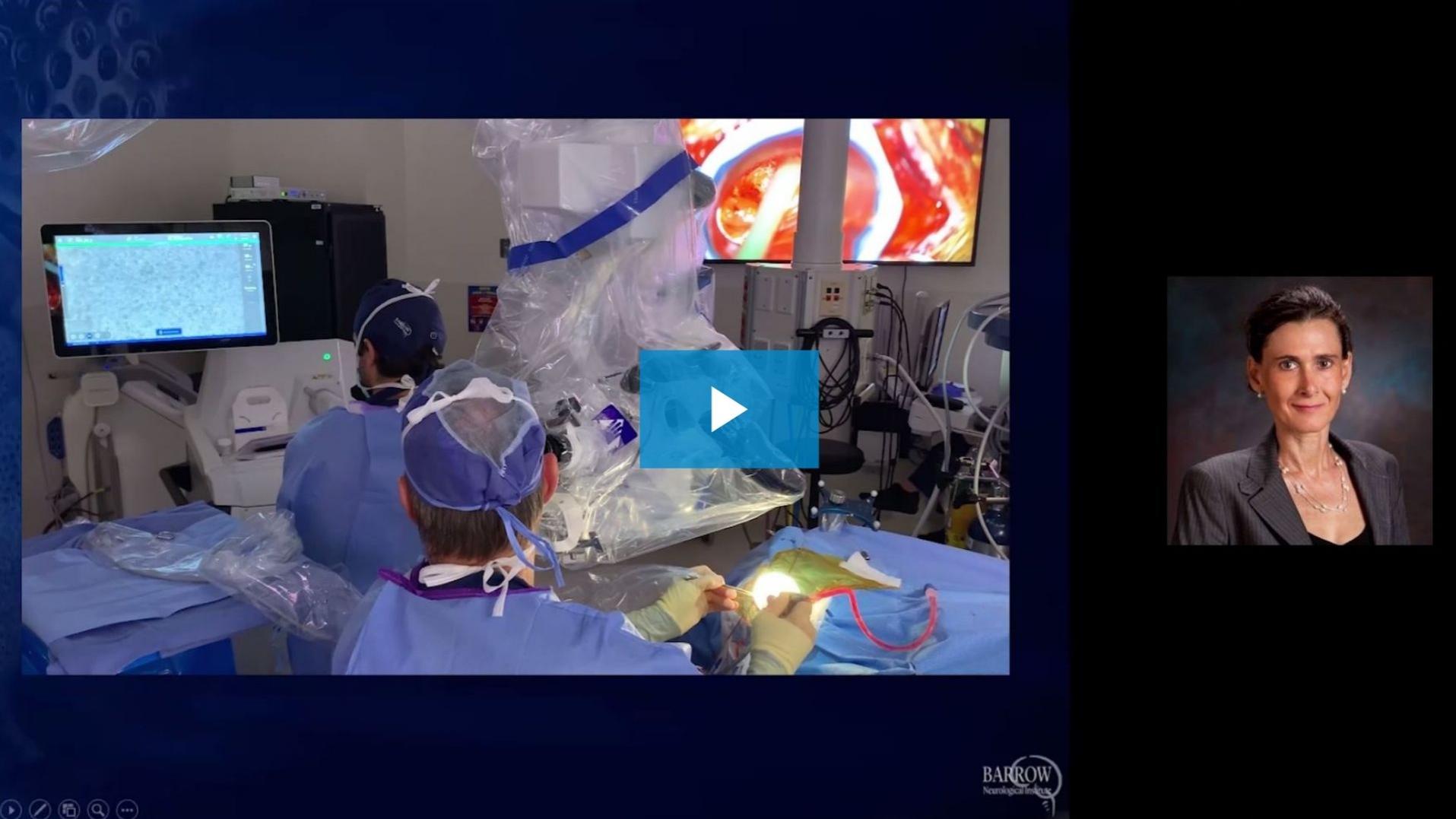
Jennifer Eschbacher, MD, partilha a sua experiência com biópsias virtuais de tumores cerebrais ao nível celular obtidas com a tecnologia de endomicroscopia confocal a laser (CLE). Na sua apresentação, fala dos resultados do estudo de CLE em curso. Além disso, fornece informações sobre um caso de anatomia patológica in vivo gravado, que foi seguido remotamente a partir do laboratório de patologia, e demonstra a interação da anatomia patológica em neurocirurgia.
![]({"xsmall":"https://www.zeiss.com/content/dam/med-hcp/reference-master/myzeiss/peer-insights/free/pathology/642-correlation-of-intraoperative-cle-images-and-conventional-histopathology/642--correlation-of-intraoperative-cle-images-and-conventional-h.jpg/_jcr_content/renditions/original.image_file.100.56.0,2,1916,1079.file/642--correlation-of-intraoperative-cle-images-and-conventional-h.jpg","small":"https://www.zeiss.com/content/dam/med-hcp/reference-master/myzeiss/peer-insights/free/pathology/642-correlation-of-intraoperative-cle-images-and-conventional-histopathology/642--correlation-of-intraoperative-cle-images-and-conventional-h.jpg/_jcr_content/renditions/original.image_file.360.203.0,2,1916,1079.file/642--correlation-of-intraoperative-cle-images-and-conventional-h.jpg","medium":"https://www.zeiss.com/content/dam/med-hcp/reference-master/myzeiss/peer-insights/free/pathology/642-correlation-of-intraoperative-cle-images-and-conventional-histopathology/642--correlation-of-intraoperative-cle-images-and-conventional-h.jpg/_jcr_content/renditions/original.image_file.768.432.0,2,1916,1079.file/642--correlation-of-intraoperative-cle-images-and-conventional-h.jpg","large":"https://www.zeiss.com/content/dam/med-hcp/reference-master/myzeiss/peer-insights/free/pathology/642-correlation-of-intraoperative-cle-images-and-conventional-histopathology/642--correlation-of-intraoperative-cle-images-and-conventional-h.jpg/_jcr_content/renditions/original.image_file.1024.576.0,2,1916,1079.file/642--correlation-of-intraoperative-cle-images-and-conventional-h.jpg","xlarge":"https://www.zeiss.com/content/dam/med-hcp/reference-master/myzeiss/peer-insights/free/pathology/642-correlation-of-intraoperative-cle-images-and-conventional-histopathology/642--correlation-of-intraoperative-cle-images-and-conventional-h.jpg/_jcr_content/renditions/original.image_file.1280.720.0,2,1916,1079.file/642--correlation-of-intraoperative-cle-images-and-conventional-h.jpg","xxlarge":"https://www.zeiss.com/content/dam/med-hcp/reference-master/myzeiss/peer-insights/free/pathology/642-correlation-of-intraoperative-cle-images-and-conventional-histopathology/642--correlation-of-intraoperative-cle-images-and-conventional-h.jpg/_jcr_content/renditions/original.image_file.1440.810.0,2,1916,1079.file/642--correlation-of-intraoperative-cle-images-and-conventional-h.jpg","max":"https://www.zeiss.com/content/dam/med-hcp/reference-master/myzeiss/peer-insights/free/pathology/642-correlation-of-intraoperative-cle-images-and-conventional-histopathology/642--correlation-of-intraoperative-cle-images-and-conventional-h.jpg/_jcr_content/renditions/original.image_file.1915.1077.0,2,1916,1079.file/642--correlation-of-intraoperative-cle-images-and-conventional-h.jpg"})