Webinário a pedido
Experiências com endomicroscopia confocal durante a ressecção de tumores do SNC
Webinar gravado durante a ZEISS Neuro Week 2020
16 dezembro 2020
· 18 MIN VISUALIZAÇÃO

Autor
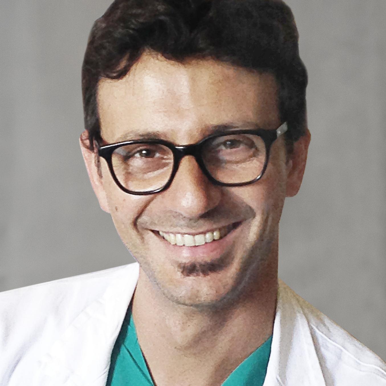
Francesco Acerbi, MD, PhD
Foundation IRCCS Istituto Neurologico Besta, Milão, Itália
RESUMO
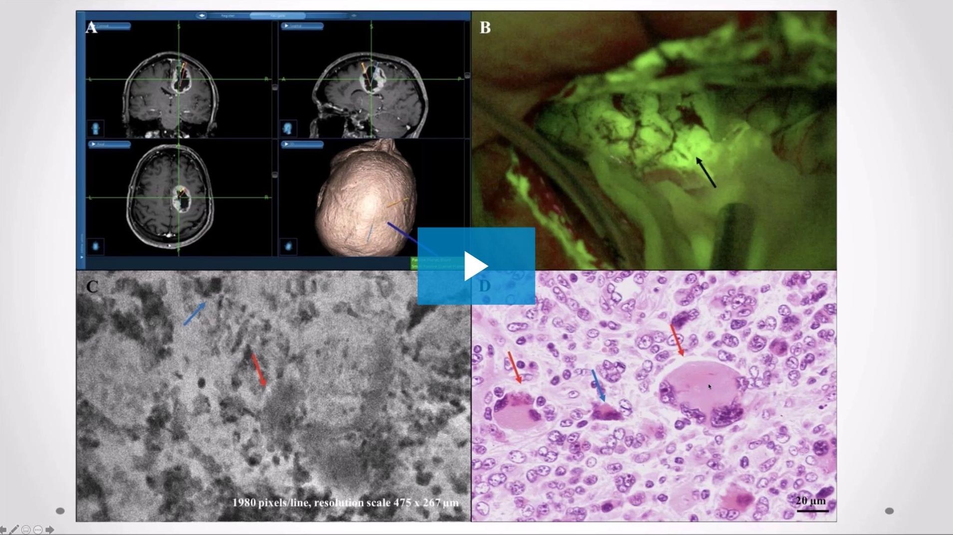
Endomicroscopia confocal guiada por fluoresceína ex vivo e in vivo durante a ressecção de tumores do SNC: experiência no Istituto Neurologico Besta
Neste webinar gravado, Francesco Acerbi, MD, PhD partilha as suas experiências clínicas com a utilização de endomicroscopia confocal guiada por fluoresceína durante a remoção de tumores do SNC. Partilha os resultados de um estudo ex vivo que compara os resultados diagnósticos e morfológicos do tecido tumoral com a utilização da microscopia confocal a laser com os resultados da secção congelada e da histologia padrão. Além disso, discute o potencial da endomicroscopia confocal para melhorar a extensão da ressecção (EOR).