Webinário a pedido
Experiências in vivo com endomicroscopia confocal
Apresentação e debate de especialistas gravada durante o primeiro CONVIVO Clinical Symposium
18 dezembro 2020
· 33 MIN VISUALIZAÇÃO
![]({"xsmall":"https://www.zeiss.com/content/dam/med-hcp/reference-master/myzeiss/peer-insights/free/pathology/509-neurosurgical-workflow-for-patients-with-brain-tumors/jennifer-eschbacher.jpg/_jcr_content/renditions/original.image_file.100.100.0,0,350,350.file/jennifer-eschbacher.jpg","small":"https://www.zeiss.com/content/dam/med-hcp/reference-master/myzeiss/peer-insights/free/pathology/509-neurosurgical-workflow-for-patients-with-brain-tumors/jennifer-eschbacher.jpg/_jcr_content/renditions/original.image_file.350.350.0,0,350,350.file/jennifer-eschbacher.jpg","medium":"https://www.zeiss.com/content/dam/med-hcp/reference-master/myzeiss/peer-insights/free/pathology/509-neurosurgical-workflow-for-patients-with-brain-tumors/jennifer-eschbacher.jpg/_jcr_content/renditions/original.image_file.350.350.0,0,350,350.file/jennifer-eschbacher.jpg","large":"https://www.zeiss.com/content/dam/med-hcp/reference-master/myzeiss/peer-insights/free/pathology/509-neurosurgical-workflow-for-patients-with-brain-tumors/jennifer-eschbacher.jpg/_jcr_content/renditions/original.image_file.350.350.0,0,350,350.file/jennifer-eschbacher.jpg","xlarge":"https://www.zeiss.com/content/dam/med-hcp/reference-master/myzeiss/peer-insights/free/pathology/509-neurosurgical-workflow-for-patients-with-brain-tumors/jennifer-eschbacher.jpg/_jcr_content/renditions/original.image_file.350.350.0,0,350,350.file/jennifer-eschbacher.jpg","xxlarge":"https://www.zeiss.com/content/dam/med-hcp/reference-master/myzeiss/peer-insights/free/pathology/509-neurosurgical-workflow-for-patients-with-brain-tumors/jennifer-eschbacher.jpg/_jcr_content/renditions/original.image_file.350.350.0,0,350,350.file/jennifer-eschbacher.jpg","max":"https://www.zeiss.com/content/dam/med-hcp/reference-master/myzeiss/peer-insights/free/pathology/509-neurosurgical-workflow-for-patients-with-brain-tumors/jennifer-eschbacher.jpg/_jcr_content/renditions/original.image_file.350.350.0,0,350,350.file/jennifer-eschbacher.jpg"})
Autor
Jennifer Eschbacher, MD
Barrow Neurological Institute, Phoenix, Arizona, EUA
RESUMO
Experiências in vivo com endomicroscopia confocal no Barrow Neurological Institute, Phoenix: perspetiva do patologista
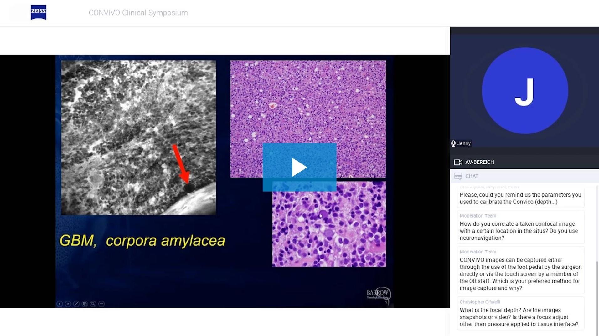
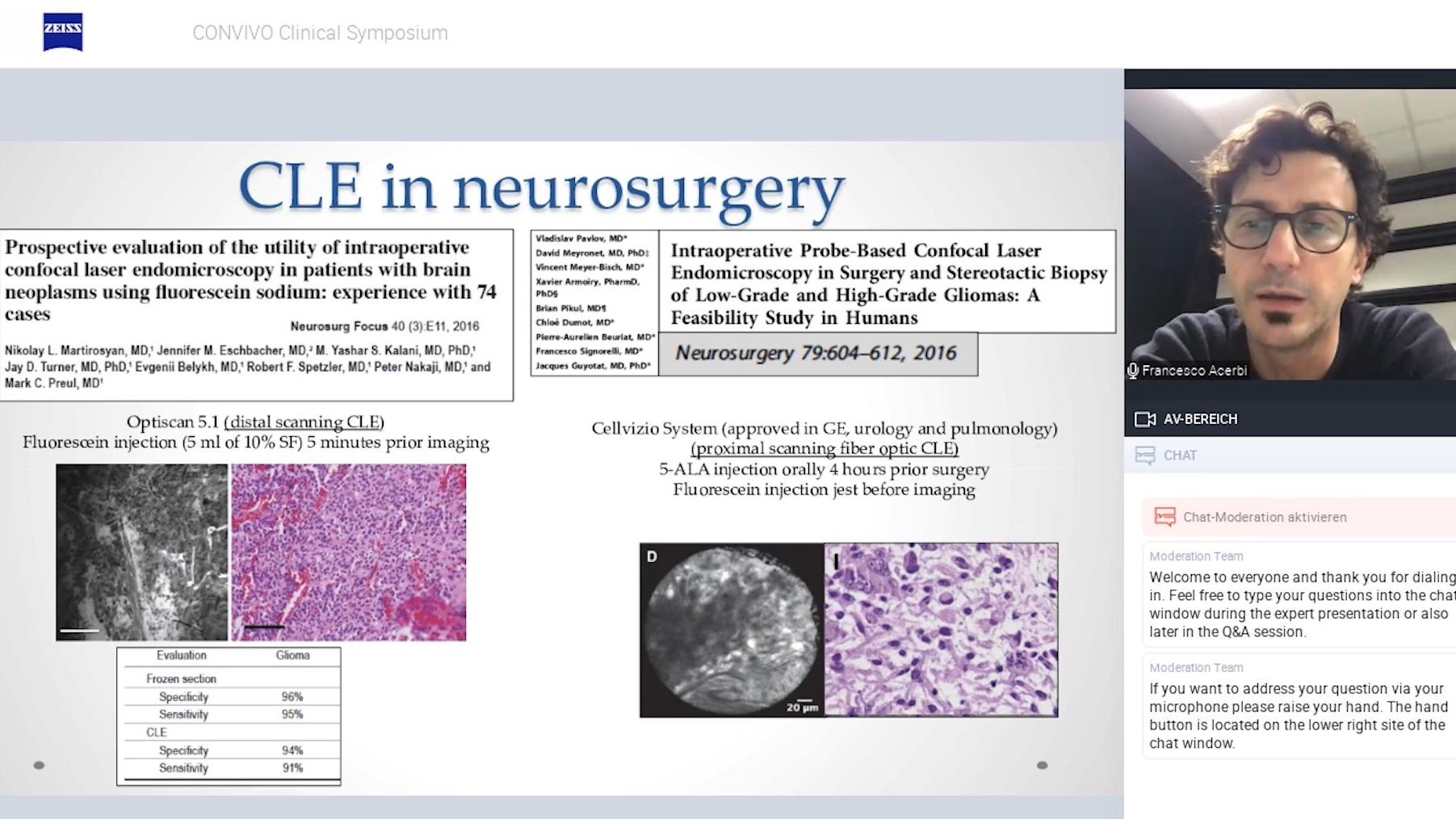
Durante este webinar gravado, a Dra. Jennifer Eschbacher partilha as suas experiências com a endomicroscopia confocal guiada por fluoresceína no Barrow Neurological Institute. Na sua palestra, discute as limitações das biópsias de secção congelada e mostra a celularidade dos pares de imagens confocais e coradas com H&E com a ajuda de diferentes exemplos de casos. Além disso, aborda as vantagens de um fluxo de trabalho de consulta de anatomia patológica in vivo para equipas médicas multifuncionais. A sua apresentação é seguida de um breve debate, durante o qual a Dra. Eschbacher responde a perguntas da audiência, fornecendo informações e orientações práticas.
![]({"xsmall":"https://www.zeiss.com/content/dam/med-hcp/reference-master/myzeiss/peer-insights/free/pathology/522-confocal-endomicroscopy-in-vivo-experiences/522-confocal-endomicroscopy-in-vivo-experiences_preview.jpg/_jcr_content/renditions/original.image_file.100.56.3,5,1916,1081.file/522-confocal-endomicroscopy-in-vivo-experiences_preview.jpg","small":"https://www.zeiss.com/content/dam/med-hcp/reference-master/myzeiss/peer-insights/free/pathology/522-confocal-endomicroscopy-in-vivo-experiences/522-confocal-endomicroscopy-in-vivo-experiences_preview.jpg/_jcr_content/renditions/original.image_file.360.203.3,5,1916,1081.file/522-confocal-endomicroscopy-in-vivo-experiences_preview.jpg","medium":"https://www.zeiss.com/content/dam/med-hcp/reference-master/myzeiss/peer-insights/free/pathology/522-confocal-endomicroscopy-in-vivo-experiences/522-confocal-endomicroscopy-in-vivo-experiences_preview.jpg/_jcr_content/renditions/original.image_file.768.432.3,5,1916,1081.file/522-confocal-endomicroscopy-in-vivo-experiences_preview.jpg","large":"https://www.zeiss.com/content/dam/med-hcp/reference-master/myzeiss/peer-insights/free/pathology/522-confocal-endomicroscopy-in-vivo-experiences/522-confocal-endomicroscopy-in-vivo-experiences_preview.jpg/_jcr_content/renditions/original.image_file.1024.576.3,5,1916,1081.file/522-confocal-endomicroscopy-in-vivo-experiences_preview.jpg","xlarge":"https://www.zeiss.com/content/dam/med-hcp/reference-master/myzeiss/peer-insights/free/pathology/522-confocal-endomicroscopy-in-vivo-experiences/522-confocal-endomicroscopy-in-vivo-experiences_preview.jpg/_jcr_content/renditions/original.image_file.1280.720.3,5,1916,1081.file/522-confocal-endomicroscopy-in-vivo-experiences_preview.jpg","xxlarge":"https://www.zeiss.com/content/dam/med-hcp/reference-master/myzeiss/peer-insights/free/pathology/522-confocal-endomicroscopy-in-vivo-experiences/522-confocal-endomicroscopy-in-vivo-experiences_preview.jpg/_jcr_content/renditions/original.image_file.1440.810.3,5,1916,1081.file/522-confocal-endomicroscopy-in-vivo-experiences_preview.jpg","max":"https://www.zeiss.com/content/dam/med-hcp/reference-master/myzeiss/peer-insights/free/pathology/522-confocal-endomicroscopy-in-vivo-experiences/522-confocal-endomicroscopy-in-vivo-experiences_preview.jpg/_jcr_content/renditions/original.image_file.1913.1076.3,5,1916,1081.file/522-confocal-endomicroscopy-in-vivo-experiences_preview.jpg"})