Webinário a pedido
Novo fluxo de trabalho neurocirúrgico para pacientes com tumores cerebrais
7 dezembro 2020
· 102 MIN VISUALIZAÇÃO
![]({"xsmall":"https://www.zeiss.com/content/dam/med-hcp/reference-master/myzeiss/peer-insights/free/pathology/509-neurosurgical-workflow-for-patients-with-brain-tumors/walavan-sivakumar.jpg/_jcr_content/renditions/original.image_file.100.100.0,10,255,265.file/walavan-sivakumar.jpg","small":"https://www.zeiss.com/content/dam/med-hcp/reference-master/myzeiss/peer-insights/free/pathology/509-neurosurgical-workflow-for-patients-with-brain-tumors/walavan-sivakumar.jpg/_jcr_content/renditions/original.image_file.255.255.0,10,255,265.file/walavan-sivakumar.jpg","medium":"https://www.zeiss.com/content/dam/med-hcp/reference-master/myzeiss/peer-insights/free/pathology/509-neurosurgical-workflow-for-patients-with-brain-tumors/walavan-sivakumar.jpg/_jcr_content/renditions/original.image_file.255.255.0,10,255,265.file/walavan-sivakumar.jpg","large":"https://www.zeiss.com/content/dam/med-hcp/reference-master/myzeiss/peer-insights/free/pathology/509-neurosurgical-workflow-for-patients-with-brain-tumors/walavan-sivakumar.jpg/_jcr_content/renditions/original.image_file.255.255.0,10,255,265.file/walavan-sivakumar.jpg","xlarge":"https://www.zeiss.com/content/dam/med-hcp/reference-master/myzeiss/peer-insights/free/pathology/509-neurosurgical-workflow-for-patients-with-brain-tumors/walavan-sivakumar.jpg/_jcr_content/renditions/original.image_file.255.255.0,10,255,265.file/walavan-sivakumar.jpg","xxlarge":"https://www.zeiss.com/content/dam/med-hcp/reference-master/myzeiss/peer-insights/free/pathology/509-neurosurgical-workflow-for-patients-with-brain-tumors/walavan-sivakumar.jpg/_jcr_content/renditions/original.image_file.255.255.0,10,255,265.file/walavan-sivakumar.jpg","max":"https://www.zeiss.com/content/dam/med-hcp/reference-master/myzeiss/peer-insights/free/pathology/509-neurosurgical-workflow-for-patients-with-brain-tumors/walavan-sivakumar.jpg/_jcr_content/renditions/original.image_file.255.255.0,10,255,265.file/walavan-sivakumar.jpg"})
Autor
Walavan Sivakumar, MD
Neurocirurgião, Pacific Neuroscience Institute, Califórnia, EUA
![]({"xsmall":"https://www.zeiss.com/content/dam/med-hcp/reference-master/myzeiss/peer-insights/free/pathology/555-advancements-in-local-brain-tumor-treatment/nakaji.png/_jcr_content/renditions/original.image_file.100.100.0,14,261,275.file/nakaji.png","small":"https://www.zeiss.com/content/dam/med-hcp/reference-master/myzeiss/peer-insights/free/pathology/555-advancements-in-local-brain-tumor-treatment/nakaji.png/_jcr_content/renditions/original.image_file.261.261.0,14,261,275.file/nakaji.png","medium":"https://www.zeiss.com/content/dam/med-hcp/reference-master/myzeiss/peer-insights/free/pathology/555-advancements-in-local-brain-tumor-treatment/nakaji.png/_jcr_content/renditions/original.image_file.261.261.0,14,261,275.file/nakaji.png","large":"https://www.zeiss.com/content/dam/med-hcp/reference-master/myzeiss/peer-insights/free/pathology/555-advancements-in-local-brain-tumor-treatment/nakaji.png/_jcr_content/renditions/original.image_file.261.261.0,14,261,275.file/nakaji.png","xlarge":"https://www.zeiss.com/content/dam/med-hcp/reference-master/myzeiss/peer-insights/free/pathology/555-advancements-in-local-brain-tumor-treatment/nakaji.png/_jcr_content/renditions/original.image_file.261.261.0,14,261,275.file/nakaji.png","xxlarge":"https://www.zeiss.com/content/dam/med-hcp/reference-master/myzeiss/peer-insights/free/pathology/555-advancements-in-local-brain-tumor-treatment/nakaji.png/_jcr_content/renditions/original.image_file.261.261.0,14,261,275.file/nakaji.png","max":"https://www.zeiss.com/content/dam/med-hcp/reference-master/myzeiss/peer-insights/free/pathology/555-advancements-in-local-brain-tumor-treatment/nakaji.png/_jcr_content/renditions/original.image_file.261.261.0,14,261,275.file/nakaji.png"})
Autor
Peter Nakaji, MD
Neurocirurgião, University of Arizona College of Medicine, EUA
![]({"xsmall":"https://www.zeiss.com/content/dam/med-hcp/reference-master/myzeiss/peer-insights/free/pathology/509-neurosurgical-workflow-for-patients-with-brain-tumors/jennifer-eschbacher.jpg/_jcr_content/renditions/original.image_file.100.100.0,0,350,350.file/jennifer-eschbacher.jpg","small":"https://www.zeiss.com/content/dam/med-hcp/reference-master/myzeiss/peer-insights/free/pathology/509-neurosurgical-workflow-for-patients-with-brain-tumors/jennifer-eschbacher.jpg/_jcr_content/renditions/original.image_file.350.350.0,0,350,350.file/jennifer-eschbacher.jpg","medium":"https://www.zeiss.com/content/dam/med-hcp/reference-master/myzeiss/peer-insights/free/pathology/509-neurosurgical-workflow-for-patients-with-brain-tumors/jennifer-eschbacher.jpg/_jcr_content/renditions/original.image_file.350.350.0,0,350,350.file/jennifer-eschbacher.jpg","large":"https://www.zeiss.com/content/dam/med-hcp/reference-master/myzeiss/peer-insights/free/pathology/509-neurosurgical-workflow-for-patients-with-brain-tumors/jennifer-eschbacher.jpg/_jcr_content/renditions/original.image_file.350.350.0,0,350,350.file/jennifer-eschbacher.jpg","xlarge":"https://www.zeiss.com/content/dam/med-hcp/reference-master/myzeiss/peer-insights/free/pathology/509-neurosurgical-workflow-for-patients-with-brain-tumors/jennifer-eschbacher.jpg/_jcr_content/renditions/original.image_file.350.350.0,0,350,350.file/jennifer-eschbacher.jpg","xxlarge":"https://www.zeiss.com/content/dam/med-hcp/reference-master/myzeiss/peer-insights/free/pathology/509-neurosurgical-workflow-for-patients-with-brain-tumors/jennifer-eschbacher.jpg/_jcr_content/renditions/original.image_file.350.350.0,0,350,350.file/jennifer-eschbacher.jpg","max":"https://www.zeiss.com/content/dam/med-hcp/reference-master/myzeiss/peer-insights/free/pathology/509-neurosurgical-workflow-for-patients-with-brain-tumors/jennifer-eschbacher.jpg/_jcr_content/renditions/original.image_file.350.350.0,0,350,350.file/jennifer-eschbacher.jpg"})
Autor
Jennifer Eschbacher, MD
Neuropatologista, Barrow Neurological Institute, Phoenix, Arizona, EUA
![]({"xsmall":"https://www.zeiss.com/content/dam/med-hcp/reference-master/myzeiss/peer-insights/free/iort/2_1607437646358/christopher-cifarelli.jpg/_jcr_content/renditions/original.image_file.100.100.0,3,255,258.file/christopher-cifarelli.jpg","small":"https://www.zeiss.com/content/dam/med-hcp/reference-master/myzeiss/peer-insights/free/iort/2_1607437646358/christopher-cifarelli.jpg/_jcr_content/renditions/original.image_file.255.255.0,3,255,258.file/christopher-cifarelli.jpg","medium":"https://www.zeiss.com/content/dam/med-hcp/reference-master/myzeiss/peer-insights/free/iort/2_1607437646358/christopher-cifarelli.jpg/_jcr_content/renditions/original.image_file.255.255.0,3,255,258.file/christopher-cifarelli.jpg","large":"https://www.zeiss.com/content/dam/med-hcp/reference-master/myzeiss/peer-insights/free/iort/2_1607437646358/christopher-cifarelli.jpg/_jcr_content/renditions/original.image_file.255.255.0,3,255,258.file/christopher-cifarelli.jpg","xlarge":"https://www.zeiss.com/content/dam/med-hcp/reference-master/myzeiss/peer-insights/free/iort/2_1607437646358/christopher-cifarelli.jpg/_jcr_content/renditions/original.image_file.255.255.0,3,255,258.file/christopher-cifarelli.jpg","xxlarge":"https://www.zeiss.com/content/dam/med-hcp/reference-master/myzeiss/peer-insights/free/iort/2_1607437646358/christopher-cifarelli.jpg/_jcr_content/renditions/original.image_file.255.255.0,3,255,258.file/christopher-cifarelli.jpg","max":"https://www.zeiss.com/content/dam/med-hcp/reference-master/myzeiss/peer-insights/free/iort/2_1607437646358/christopher-cifarelli.jpg/_jcr_content/renditions/original.image_file.255.255.0,3,255,258.file/christopher-cifarelli.jpg"})
Autor
Christopher Cifarelli, MD, PhD
Diretor do Programa de Radiocirurgia Gamma Knife da Radiation Oncology da West Virginia University, EUA
![]({"xsmall":"https://www.zeiss.com/content/dam/med-hcp/reference-master/myzeiss/peer-insights/free/neurosurgery/1_1608037835433/klaus-henning-kahl.jpg/_jcr_content/renditions/original.image_file.100.100.0,31,255,286.file/klaus-henning-kahl.jpg","small":"https://www.zeiss.com/content/dam/med-hcp/reference-master/myzeiss/peer-insights/free/neurosurgery/1_1608037835433/klaus-henning-kahl.jpg/_jcr_content/renditions/original.image_file.255.255.0,31,255,286.file/klaus-henning-kahl.jpg","medium":"https://www.zeiss.com/content/dam/med-hcp/reference-master/myzeiss/peer-insights/free/neurosurgery/1_1608037835433/klaus-henning-kahl.jpg/_jcr_content/renditions/original.image_file.255.255.0,31,255,286.file/klaus-henning-kahl.jpg","large":"https://www.zeiss.com/content/dam/med-hcp/reference-master/myzeiss/peer-insights/free/neurosurgery/1_1608037835433/klaus-henning-kahl.jpg/_jcr_content/renditions/original.image_file.255.255.0,31,255,286.file/klaus-henning-kahl.jpg","xlarge":"https://www.zeiss.com/content/dam/med-hcp/reference-master/myzeiss/peer-insights/free/neurosurgery/1_1608037835433/klaus-henning-kahl.jpg/_jcr_content/renditions/original.image_file.255.255.0,31,255,286.file/klaus-henning-kahl.jpg","xxlarge":"https://www.zeiss.com/content/dam/med-hcp/reference-master/myzeiss/peer-insights/free/neurosurgery/1_1608037835433/klaus-henning-kahl.jpg/_jcr_content/renditions/original.image_file.255.255.0,31,255,286.file/klaus-henning-kahl.jpg","max":"https://www.zeiss.com/content/dam/med-hcp/reference-master/myzeiss/peer-insights/free/neurosurgery/1_1608037835433/klaus-henning-kahl.jpg/_jcr_content/renditions/original.image_file.255.255.0,31,255,286.file/klaus-henning-kahl.jpg"})
Autor
Henning Kahl, MD
Radioncologista, University Hospital, Augsburgo, Alemanha
RESUMO
Perspetivas de um novo fluxo de trabalho neurocirúrgico para pacientes com tumores cerebrais
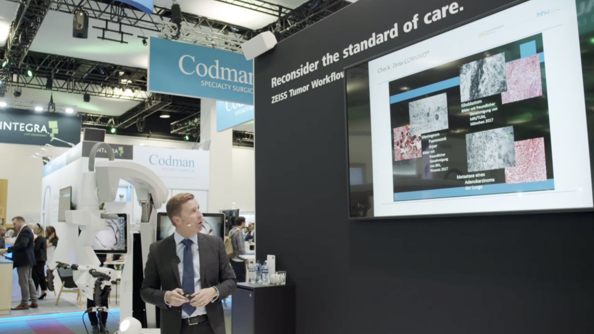
Durante este webinar multidisciplinar da ZEISS, neurocirurgiões, radioncologistas e uma neuropatologista partilham as suas ideias sobre a aplicação das tecnologias do ZEISS Tumor Workflow.
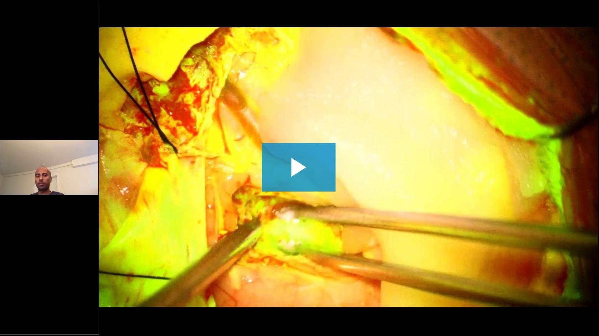
São abordados os seguintes tópicos: a utilização de fluoresceína para tumores cerebrais, experiências clínicas com o sistema de endomicroscopia confocal in vivo ZEISS CONVIVO do ponto de vista neurocirúrgico (a partir do minuto 24:05) e neuropatológico (a partir do minuto 37:00), bem como a perspetiva dos radioncologistas (a partir do minuto 1:01:00) sobre o papel da IORT no tratamento de metástases cerebrais e glioblastoma.
![]({"xsmall":"https://www.zeiss.com/content/dam/med-hcp/reference-master/myzeiss/peer-insights/free/pathology/509-neurosurgical-workflow-for-patients-with-brain-tumors/509-neurosurgical-workflow-for-patients-with-brain-tumors_preview.jpg/_jcr_content/renditions/original.image_file.100.56.6,5,1919,1081.file/509-neurosurgical-workflow-for-patients-with-brain-tumors_preview.jpg","small":"https://www.zeiss.com/content/dam/med-hcp/reference-master/myzeiss/peer-insights/free/pathology/509-neurosurgical-workflow-for-patients-with-brain-tumors/509-neurosurgical-workflow-for-patients-with-brain-tumors_preview.jpg/_jcr_content/renditions/original.image_file.360.203.6,5,1919,1081.file/509-neurosurgical-workflow-for-patients-with-brain-tumors_preview.jpg","medium":"https://www.zeiss.com/content/dam/med-hcp/reference-master/myzeiss/peer-insights/free/pathology/509-neurosurgical-workflow-for-patients-with-brain-tumors/509-neurosurgical-workflow-for-patients-with-brain-tumors_preview.jpg/_jcr_content/renditions/original.image_file.768.432.6,5,1919,1081.file/509-neurosurgical-workflow-for-patients-with-brain-tumors_preview.jpg","large":"https://www.zeiss.com/content/dam/med-hcp/reference-master/myzeiss/peer-insights/free/pathology/509-neurosurgical-workflow-for-patients-with-brain-tumors/509-neurosurgical-workflow-for-patients-with-brain-tumors_preview.jpg/_jcr_content/renditions/original.image_file.1024.576.6,5,1919,1081.file/509-neurosurgical-workflow-for-patients-with-brain-tumors_preview.jpg","xlarge":"https://www.zeiss.com/content/dam/med-hcp/reference-master/myzeiss/peer-insights/free/pathology/509-neurosurgical-workflow-for-patients-with-brain-tumors/509-neurosurgical-workflow-for-patients-with-brain-tumors_preview.jpg/_jcr_content/renditions/original.image_file.1280.720.6,5,1919,1081.file/509-neurosurgical-workflow-for-patients-with-brain-tumors_preview.jpg","xxlarge":"https://www.zeiss.com/content/dam/med-hcp/reference-master/myzeiss/peer-insights/free/pathology/509-neurosurgical-workflow-for-patients-with-brain-tumors/509-neurosurgical-workflow-for-patients-with-brain-tumors_preview.jpg/_jcr_content/renditions/original.image_file.1440.810.6,5,1919,1081.file/509-neurosurgical-workflow-for-patients-with-brain-tumors_preview.jpg","max":"https://www.zeiss.com/content/dam/med-hcp/reference-master/myzeiss/peer-insights/free/pathology/509-neurosurgical-workflow-for-patients-with-brain-tumors/509-neurosurgical-workflow-for-patients-with-brain-tumors_preview.jpg/_jcr_content/renditions/original.image_file.1913.1076.6,5,1919,1081.file/509-neurosurgical-workflow-for-patients-with-brain-tumors_preview.jpg"})