3D X-ray Imaging in Life Science Research
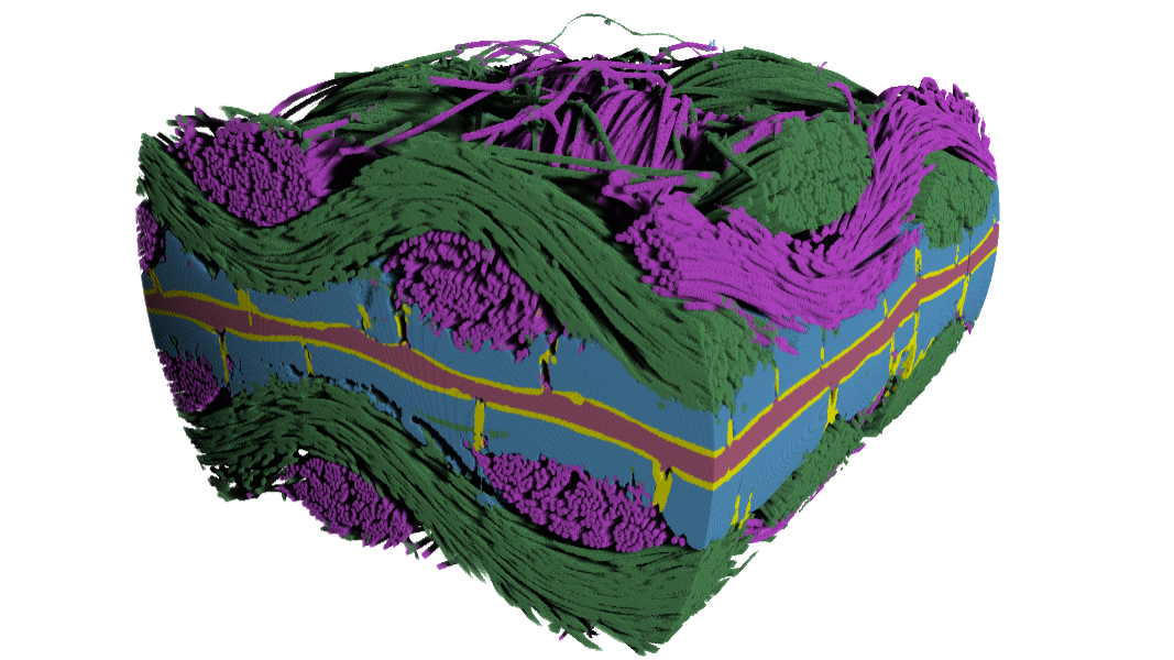
Webinar | 27 June @ 2 pm CESTUnderstanding physiological structure is at the core of many research questions in life science. Fluorescence microscopy is used to visualize specific, labelled structures and electron microscopy offers ultraresolution information of smaller regions. However, life scientists with a need to better understand anatomical structure in bulk samples have been increasingly turning to X-rays for their imaging needs. X-ray tomography captures larger volumes of structural information down to sub-micron resolution from samples ranging from millimeters to tens of centimeters in size. Specimens as varied as plant tissues, whole organisms, organs, mineralized and soft tissues can be imaged, and this approach is becoming a key technology for life scientists who need a broader picture of the anatomical structures they are investigating. Use X-ray tomography to study the internal histology of your specimens down to a cellular level, without destroying your sample with dissection.