XRM Webinar Series

X-ray Microscopy and Progress in Materials Science Join our Webinar | 21 March 2024

  • Acquire insights and best practices of materials characterization by means of X-ray microscopes as a stand alone technique or a part of a correlative workflow
  • Gain practical knowledge and understanding on the potential of X-ray microscopy in materials characterization
  • Get information on the recent achievements in the field of materials sciences using X-ray microscopes

Welcome to the 1st episode of our upcoming quarterly webinar series

Segmented 3D volume of a polymer electrolyte fuel cell membrane electrode assembly. Segmented 3D volume of a polymer electrolyte fuel cell membrane electrode assembly. Gas diffusion layer fiber weaves are visible in green and magenta, microporous layer in blue, catalyst in yellow, and electrolyte membrane in red.
Segmented 3D volume of a polymer electrolyte fuel cell membrane electrode assembly. Gas diffusion layer fiber weaves are visible in green and magenta, microporous layer in blue, catalyst in yellow, and electrolyte membrane in red.
 Segmented 3D volume of a polymer electr olyte fuel cell membrane electrode assembly. Gas diffusion layer fiber weaves are visible in green and magenta, microporous layer in blue, catalyst in yellow, and electrolyte membrane in red.
 Segmented 3D volume of a polymer electr olyte fuel cell membrane electrode assembly. Gas diffusion layer fiber weaves are visible in green and magenta, microporous layer in blue, catalyst in yellow, and electrolyte membrane in red.
Segmented 3D volume of a polymer electr olyte fuel cell membrane electrode assembly. Gas diffusion layer fiber weaves are visible in green and magenta, microporous layer in blue, catalyst in yellow, and electrolyte membrane in red.

X-ray Microscopy and Progress in Materials Science

Online Webinar | 21 March @ 2 pm CET

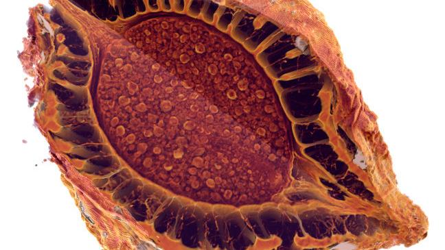
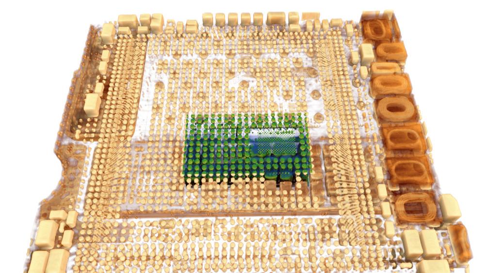
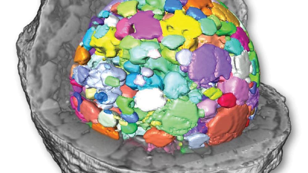
X-ray microscopy has established itself as a key characterization method in the field of Materials Science owing to the 3D non-destructive imaging capabilities of the technique. These qualities enable deep insights into the microstructures of materials in their native states and unlock the potential for observing microstructural evolutions due to, e.g., processing, mechanical loading, or environmental exposure. This webinar highlights advancements in the field of x-ray microscopy enabled by new developments in instrumentation hardware and machine learning assisted reconstruction software and demonstrate recent application examples across the diverse disciplines covered by Materials Science.

Webinar Speakers

Webinar Presenter: Mohsen Samadi Khoshkhoo
Presenter Dr. Mohsen Samadi Khoshkhoo

Mohsen Samadi Khoshkhoo is a Business Development Manager at ZEISS for X-ray microscopy systems in the region Europe, the Middle East and Africa (EMEA). With his BSc and MSc in metallurgy he worked for a year in a steel company followed by a year in a materials and energy research center in Iran. In 2009 he joined to Leibniz Institute for Solid State and Materials Research in Dresden (IFW Dresden) and TU Dresden, where he conducted his PhD in Materials Science. Materials characterization was the core of his work at IFW where he dealt with scanning electron microscopy (SEM), transmission electron microscopy (TEM), in-situ SEM, X-ray line profile analysis, and time resolved synchrotron X-ray diffraction investigations. He has been a member of the ZEISS team since 2014.

Webinar Presenter: Stephen Kelly
Presenter Stephen Kelly

Stephen Kelly is the Market Sector Manager for Energy Materials at Carl Zeiss RMS. He has been working in the battery and energy materials space for over 20 years and has extensive experience with materials characterization and fabrication across the energy space. His expertise covers batteries, fuel cells, hydrogen storage materials, and photovoltaics, among others. After receiving his BS in Engineering Physics from Colorado School of Mines in 2002, he went on to receive his Ph.D. in Materials Science and Engineering from Stanford University in 2009. He spent 4 years working as a postdoc at Lawrence Berkeley National Laboratory specializing in x-ray microscopy of solar cells and atmospheric aerosols. He has been working at Carl Zeiss RMS for the last 7 years as an imaging specialist and Sector Manager for the Energy Materials market. He lives in San Francisco, California.

Watch the webinar recording

Don´t miss out our next Webinar sessions on XRM

Here is what´s about to come soon