Product image of ZEISS LSM Lightfield 4D
Product

LSM Lightfield 4D

Instant Volumetric High-Speed Imaging of Living Organisms

Lightfield 4D is instant volumetric imaging at high speed. Acquire comprehensive 3D information with a single snap and say goodbye to any time delay within an imaged volume. For the first time, capture the fastest movements within whole organisms at up to 80 volumes per second – with all spatiotemporal information intact. Crawling larvae, beating hearts, flowing blood, and firing neurons can be studied in 3D at unprecedented speed to unravel the secrets of life.

  • One snap. One volume.
  • Minimum light exposure. Maximum information gain.
  • Fast acquisition. Increased throughput.
  • One imaging platform. Endless possibilities.

One Snap. One Volume.

Capture Spatial Signals and Fast Dynamics Without Compromise

Life moves. Many neuronal and physiological processes occur at very high speeds, making it difficult to accurately capture their spatiotemporal dynamics. Although established technologies have become faster, the required acquisition time still increases with sample volume, so fast processes like neuronal activity or heartbeats require a trade-off between volumetric information and image frame rate. With Lightfield 4D, you no longer have to compromise, as you can capture 80 volumes per second without time delay in 3D. This makes it possible to follow neuronal activity in zebrafish brains, track tissue movement in developing Drosophila embryos, and keep track of moving structures in crawling C. elegans larvae. The unique one-snap-one-volume imaging ensures that crucial events are not missed or distorted. Highly time-resolved particle tracking in complete volumes is finally possible. Start your experiments immediately – on your confocal and without the need to adjust sample preparation.

  • The movie shows 3 full heartbeats in 1.2 seconds, during which cardiomyocytes are temporally and spatially resolved

    Investigating the morphology and cardiac wall movement of the developing zebrafish heart

  •  Zebrafish blood flow

    Observing red blood cells flowing through the tail vasculature of the developing zebrafish embryo in real time

  • Flow insect blood cells in the hemolymph of Drosophila melanogaster white pre-pupae

    Investigating the flow of insect blood cells (hemocytes) in the Drosophila hemolymph

  • Sensitive 3D imaging of Drosophila muscle activation

    Sensitive 3D imaging of Drosophila muscle activation

  • Transgenic, 3-day old Arabidopsis thaliana hypocotyl

    Capturing light-induced redistribution of proteins in Arabidopsis thaliana


  • Investigating the morphology and cardiac wall movement of the developing zebrafish heart

    Analyzing embryonic heart morphology and movement in 3D is challenging as the heart is continually beating. The data was recorded from a 3 days post fertilization zebrafish larvae embedded in agarose. ZEISS Lightfield 4D allowed to image the heartbeat with 80 volumes per second. The movie shows 3 full heartbeats in 1.2 seconds, during which cardiomyocytes are temporally and spatially resolved. This allows for cell segmentation and tracking using ZEISS arivis Pro. It is clearly visible that the cardiomyocytes follow exactly the same trajectory in every heartbeat.

    Courtesy of Stone Elworthy and Emily Noël, School of Biosciences, University of Sheffield, UK. Data acquired at Wolfson Light Microscopy Facility in the School of Biosciences at the University of Sheffield.

  • Observing red blood cells flowing through the tail vasculature of the developing zebrafish embryo in real time

    Imaging the behavior of the circulatory system in real time in three dimensions is far not possible, as the heart pumps blood through the circulatory system at a speed of 3 beats per second, giving cells too fast of speeds to capture their movement in more than 2 dimensions at a time. Lightfield 4D with speeds up to 80 volumes per second offer the first opportunity to track blood cells throughout this network in 3D and visualize just exactly how this network is organized with respect to relative speed and directionality of blood in different vessels throughout the volume.

    Courtesy of Toby Andrews and Rashmi Priya, The Francis Crick Institute, London, UK

  • Investigating the flow of insect blood cells (hemocytes) in the Drosophila hemolymph

    Investigating flow of hemocytes, the insect blood cells, through the hemolymph in vivo was almost impossible for researchers due to the fast three-dimensional movement. Lightfield 4D offers the unique opportunity to image a large volume fast enough to follow this process under physiological in vivo conditions. Thanks to the unparalleled imaging speed of 80 volumes per second, cells are reliably resolved both spatially and temporally. The acquired data allow subsequent segmentation and automated tracking using ZEISS arivis Pro.

    Courtesy of Iwan Robert Evans, University of Sheffield, UK. Data acquired at Wolfson Light Microscopy Facility in the School of Biosciences at the University of Sheffield.

  • Sensitive 3D imaging of Drosophila muscle activation

    Drosophila melanogaster 1st instar larvae, semi-constrained, expressing Calcium sensitive fluorescent proteins in all body wall muscles. Analysis with ZEISS arivis Pro to visualize muscle activity.

    Courtesy of Sean Sweeney, Department of Biology, University of York, UK

  • Capturing light-induced redistribution of proteins in Arabidopsis thaliana

    Investigating how plants respond to light appropriately on the molecular level is important for understanding how they carry out many of their light-dependent functions. In this situation, a re-localization of a mobile Arabidopsis thaliana protein upon blue-light stimulation is observed in the hypocotyl (stem) of a 3-day old seedling. The molecular dynamics are too fast for traditional confocal imaging. The high speed of Lightfield 4D enables an entire 3D volume to be imaged in a single snap, perfectly preserving the spatial localizations of protein aggregates. This provides reliable positioning information, which enables accurate reconstructions.

    Courtesy of Hannah Walters, Cellular Analysis Facility, MVLS-Shared Research Facilities, University of Glasgow. Data acquired at the Cellular Analysis Facility, University of Glasgow, UK
image of LSM Lightfield 4D product flyer

ZEISS LSM Lightfield 4D

Instant Volumetric High-Speed Imaging of Living Organisms

Minimum Light Exposure. Maximum Information Gain.

Observe Entire Organisms for as Long as You Want Without Altering the Processes of Life.

Collecting 3D information of living samples has always been a challenge, especially for large sample volumes. Optical sectioning requires sequential acquisition of single images to create a Z-stack. Each slice requires light exposure, which is not fully limited to the plane of illumination and can easily add up to harmful amounts across the volume. Lightfield 4D works differently: A complete Z-stack is acquired with a single illumination event, reducing light exposure and phototoxic effects to a minimum. Living samples can be imaged over long periods of time at high temporal density. This combination of outstanding 3D imaging speed and extreme gentleness allows you to follow the sample in multicolor over time without influencing the recorded living activity. You can observe developmental processes, cell migration, vesicle movement or other changes in tissues and organisms that take hours or even days to complete, and still achieve the temporal resolution needed to understand the dynamics.

  • Developing Drosophila pupa

    Observing the formation of fat body tissue in a developing Drosophila pupa

  • Zebrafish ear undergoing developmental morphogenesis

    Zebrafish ear undergoing developmental morphogenesis

  • Following gut development in Drosophila embryos

    Following gut development in Drosophila embryos


  • Observing the formation of fat body tissue in a developing Drosophila pupa

    Visualizing development of tissues and organs in intact animals enables better understanding of factors involved in their regulation and dysfunction. One example is the developing fat body forming during the pupal stage of Drosophila. Lightfield 4D allows to keep pace with the cell movement, to provide robust data for 4D tracking. Illumination is gentle enough to image overnight without sacrificing organism viability or fluorophore strength.

    15-hour overnight imaging including 12 positions and 10 animals, 500 ms exposure times per volume with 2-minute intervals.

    Courtesy of Ignacio Manuel Fernández Guerrero, Cellular Analysis Facility, MVLS-Shared Research Facilities, University of Glasgow. Data acquired at the Cellular Analysis Facility, University of Glasgow

  • Zebrafish ear undergoing developmental morphogenesis

    Morphogenesis of developing organs requires a complex coordination of varying regulators and genomic elements. Lightfield 4D enables acquisition of light-sensitive processes with sufficient resolution to track epithelial cell morphological patterning. Its one-snap-one-volume imaging not only ensures that no developmental processes are missed or lost amidst the z-stacks but even allows multiple animals to be imaged in batch mode to capture all events and increase experimental throughput.

    Zebrafish embryo, timelapse movie of developing otic vesicle, 2 – 3 days post fertilization. Every 2 minutes, volumes of 4 different zebrafish embryo ears were imaged over the course of 16 hours.

    Courtesy of Tanya Whitfield, Sarah Baxendale, School of Biosciences, University of Sheffield, UK. Data acquired at the Wolfson Light Microscopy Facility, University of Sheffield.

  • Following gut development in Drosophila embryos

    The Drosophila embryonic midgut is particularly challenging to live image due to its depth combined with the different refractive indices of the eggshell and surrounding tissues. The current practice is to either use multiphoton microscopy and risk phototoxicity, or to image mutants in which the refractive index of the embryo is shifted and risk biological side effects during characterization.

    Lightfield 4D allows to image the formation of the midgut throughout much of embryogenesis without any risk of phototoxicity, and with unparalleled clarity. The dataset was recorded for close to 7 hours with 1 volume every 10 seconds.

    Courtesy of Andrew T Plygawko, School of Biosciences, University of Sheffield, UK. Data acquired at the Wolfson Light Microscopy Facility, University of Sheffield.

Fast Acquisition. Increased Throughput.

Examine Multiple Positions or Numerous Samples with Instantaneous Volumetric Imaging.

Typically, acquisition time of large volumes is the critical factor that limits the throughput of imaging. Acquiring a large volume with a single image snap speeds up your experiments by multitudes. The unmatched speed with which Lightfield 4D captures multi-color volumes can be used to increase the productivity of experiments in a variety of ways: Image and analyze more samples than ever before in every session, immediately improving experimental statistics. Compare multiple different sample cohorts of wild type and genetically modified phenotypes, or samples with different drug treatments. Instead of hours, only minutes are spent collecting the data you need, leaving you more time for advanced analysis and investigation of your datasets.

  • Cleared spheroid

    Efficient volume imaging of cleared spheroids with subsequent cell counting

  • Imaging cancer organoids

    Imaging cancer organoids with high speed to enable assessment of perturbations


  • Efficient volume imaging of cleared spheroids with subsequent cell counting

    Traditional acquisition methods like confocal point scanning or the use of spinning disk systems take considerable time for the acquisition of z-stacks. The speed of Lightfield 4D imaging enables advanced screening applications where higher throughput is required and facilitates faster screening of many spheroids under similar and different conditions, like in compound screens and drug treatments.

    Cleared spheroid of a co-culture of HCT-116-GFP (colon cancer) / NIH-3T3-RFP (fibroblasts) cells stained with Hoechst for nuclei. Imaged in an InSphero Akura plate. Dataset was segmented using arivis Pro.

    Sample courtesy of InSphero AG. Schlieren, Switzerland

  • Imaging cancer organoids with high speed to enable assessment of perturbations

    Organoids are popular biological models for analyzing properties within cancer systems, such as responses to drug treatments, extracellular environments, and immune cell interactions. Image acquisition of such large 3D structures and screening through large sample sets is particularly time consuming. Lightfield 4D enables 3D image acquisition of organoids at a rate of several per second, dramatically increasing the throughput for screening through large numbers compared to traditional microscopy methods.

    Colorectal cancer organoids, actin cytoskeleton labelled with phalloidin (magenta), nuclei labelled with DAPI (blue). Captured with a 40× objective using 100 ms exposure time for each fluorophore.

    Courtesy of Nikki R. Paul, Cancer Research UK Scotland Institute, Glasgow. Data acquired at the Cellular Analysis Facility, University of Glasgow.

One Imaging Platform. Endless Possibilities.

Be Innovative in Your Experiments and Combine Light-Field Microscopy with All the Possibilities of an LSM.

Laser scanning microscopes (LSM) have proven to be the most versatile microscopy systems. They combine super-resolution and spectral imaging with high-quality optical sectioning of large samples, along with the capability to incorporate additional fluorescent information and molecular dynamics measurements. Take your experiments to the next level by pairing this remarkable flexibility with the gentle and instant volume imaging of Lightfield 4D:

  • Monitor neuronal activity in 3D at high speed, and supplement this with super-resolution structural details captured with Airyscan.
  • Track macrophage movement during a wound healing assay and add high-resolution details of the wound site to your investigation.
  • Leverage the photomanipulation capabilities of your LSM for bleaching, photoactivation, photoconversion, or ablation experiments, followed by gentle volume imaging.
  • Accomplish all of this on the same microscope as part of the same experiment without ever moving your sample.

The thinking zebrafish: Analyzing neuronal activity in developing organisms

Imaging calcium signaling as proxy for neuron activity is a widely used technique in many model systems. These signals occur rapidly, in milliseconds, requiring high temporal resolution.

The video shows calcium signaling in the zebrafish brain. Thanks to the large volume and speed of Lightfield 4D, neurons more than 50 µm apart from each other can be recorded at the same time. Additional high-resolution data was acquired using the Airyscan CO-8Y mode.

Data recorded from a zebrafish larvae 4 days post fertilization expressing the calcium reporter GCaMP6; image volume: 361 × 361 × 109 µm³; 10 volumes per second for 1 minute (661 time points); exposure time 91 ms; intensity coding LUT (low intensity blue, high intensity red to white).

Courtesy of Anton Nikolaev, University of Sheffield, UK. Data acquired at Wolfson Light Microscopy Facility in the School of Biosciences at the University of Sheffield.

Combine Light-Field Microscopy with LSM Flexibility

Lightfield 4D is available with ZEISS LSM 910 and ZEISS LSM 990 on ZEISS Axio Observer.

Light-Field Microscopy by ZEISS

Technology Insights

To truly capture the essence of biological processes, imaging must be done in 4D, as both volume and time are essential for investigating living systems. This concept is not new; many optical sectioning techniques have been developed over the past decades to attempt to meet this requirement. However, these methods typically rely on sequential image acquisition to create Z-stack images of volumes, which introduces time differences within the sample volume, severely limiting the imaging speed and the spatiotemporal accuracy of the acquired data.

Lightfield 4D offers a unique solution by imaging an entire volume at an exact point in time, without any time delay. Instead of capturing single 2D images at different time points, a micro lens array positioned in between objective and camera generates 37 individual images, collecting all of the 3D information at the same instant. Each of these different views provides both spatial and angular information which serves as the foundation for creating a Z-stack through deconvolution-based processing. In this way, Lightfield 4D can generate 80 volume Z-stacks per second.
 

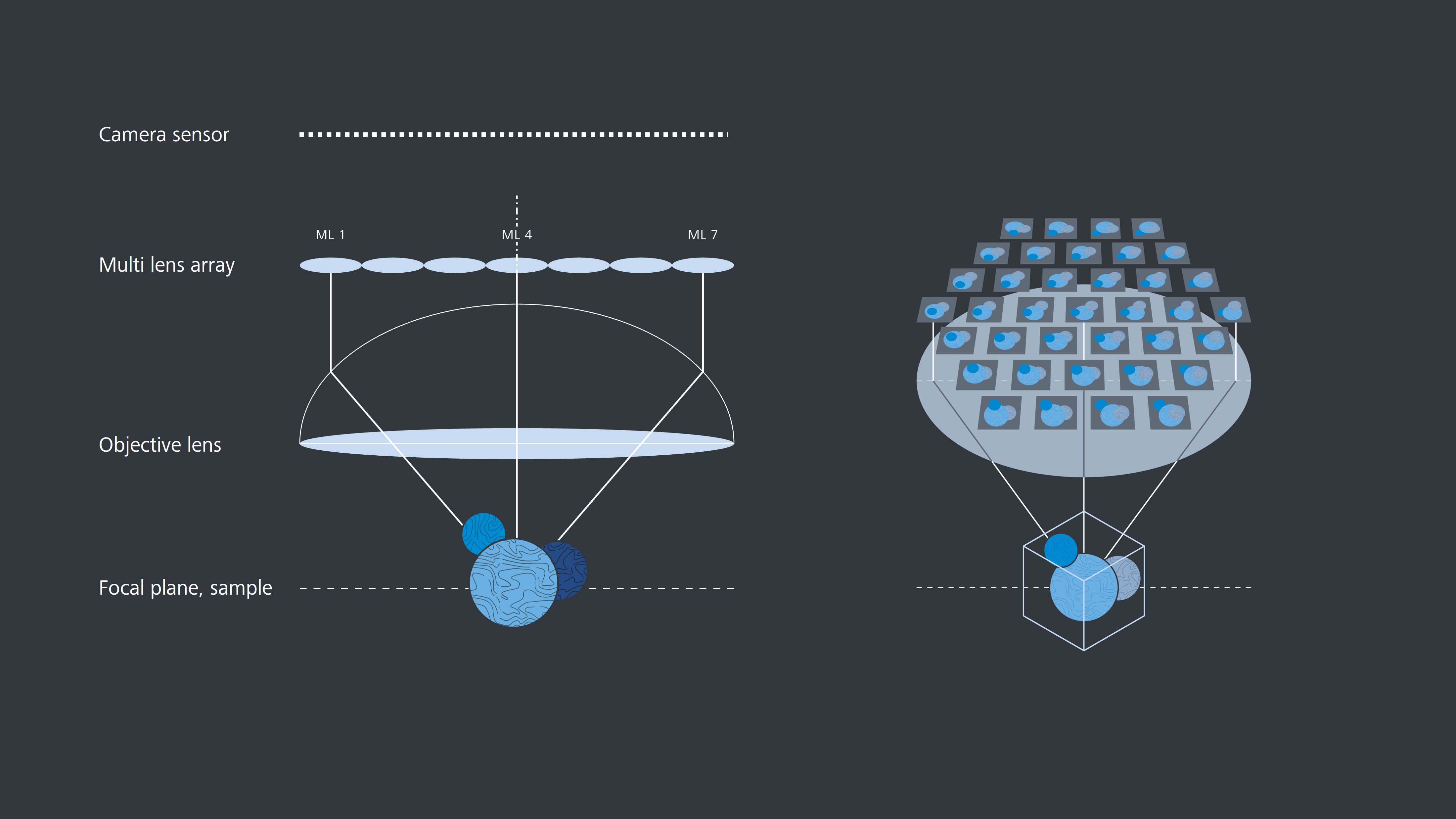
A multi lens array positioned in between objective and camera generates 37 individual images, collecting all of the 3D information at the same instant.

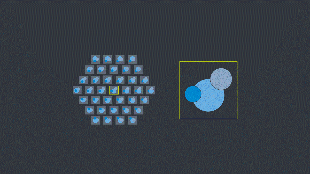
Each of 37 different views provides both spatial and angular information which contributes to the volumetric information of the sample. Lightfield 4D can generate 80 of such volumes per second.

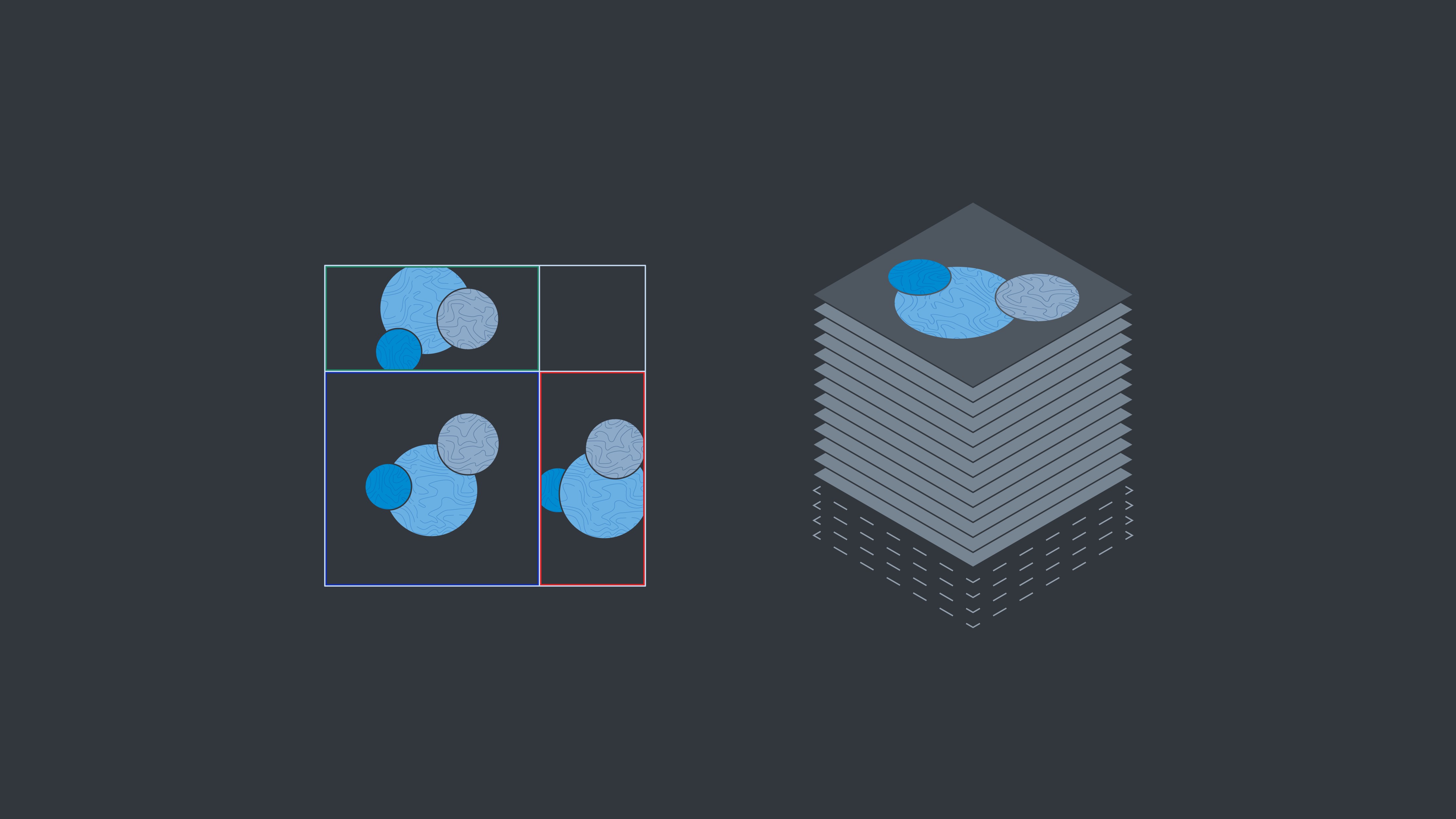
Through deconvolution-based processing, Z-stacks are generated and saved in the .czi file format, allowing for all rendering and analysis options available in ZEN and arivis Pro.

Download Your Flyer Now

image of LSM Lightfield 4D product flyer

Download the product flyer and get more information about ZEISS LSM Lightfield 4D:

  • Application examples
  • Technology insights
  • Technical specifications

Form is loading...

If you want to have more information on data processing at ZEISS please refer to our data privacy notice.