

ZEISS arivis Pro
Your Solution for Advanced Image Analysis and Visualization
Maximize your image potential with our powerful image analysis and visualization tools. Create seamless analysis pipelines with just a few clicks. Effortlessly process massive datasets on compatible workstations. “Walk into your sample” using our VR toolkit for a new perspective.


Flexible, End-to-End Image Analysis Pipelines
ZEISS arivis Pro empowers you to automate image analysis and visualization pipelines. Leverage traditional methods or AI models effortlessly to create pipelines for any image size, dimension, or modality without the need to code.
The software supports and handles over 30 commercial file formats. Efficiently process large files with ease. Pre-configured pipelines and standard assays are available for both simple and demanding analysis tasks. Or you can customize pipelines for your specific goals.
It takes just one click to repeat your analysis for consistent, quantitative results. Boost productivity and ensure reproducible results.


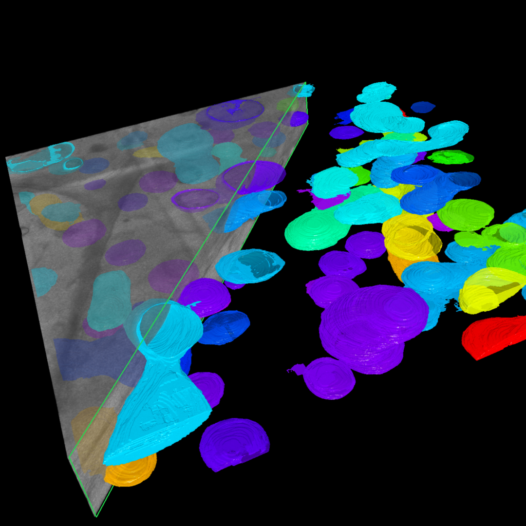
Sub cellular analysis based in AI model of FIB SEM image
Advanced 3D Analysis
Boost results quickly with AI, even for complex tasks. Annotate slices in your Z-stack to train your model segmentation and classification and then apply it to the whole dataset. Use Machine Learning and Deep Learning operators locally or train models in the cloud and then import them to form the core of your automated pipeline. Visualize results in 3D and create videos for a polished analysis report with a few clicks.


3D organoid analysis
High Content Analysis
Complex disease drug discovery demands advanced image analysis methods such as screening against a broader view of cell or tissue phenotype, scalable from 2D to 4D. Train an AI model using ZEISS arivis software on the cloud or locally, and automate image analysis to save time, reduce human bias, and gain consistent results.


Tracking with ZEISS arivis Pro VR


Tracking and Lineage
Quantify cell division and migratory phenotypes, and track changes in 2D and 3D image sets of any size. Use ZEISS arivis Pro VR to accurately proofread automatically generated tracks in an immersive VR environment. Easily reapply your pipeline to more datasets.
.")
.")
Automated 3D neuron tracing of neuronal tissue (mouse brain).
Neurobiology
Deploy cutting-edge neuron tracing algorithms for automatic, repeatable analysis of diverse images with a single click. Quantify neurons, glia/ microglia cells, axons, and more, even with low signal-to-noise ratio (SNR). Edit traces easily, using our semi-automatic interactive and VR tools. Analyze past experiment data, no matter the image format.


ZEISS arivis Pro VR Toolkit
Get a new perspective on your sample with an immersive virtual reality experience. Freed from being tethered to your keyboard and mouse, you can directly move, rotate, scale, and shape your digital image data. Experience an in-depth perception equivalent to the real world.
- Explore your sample in 3D
- Observe details from diverse angles
- Collaborate with colleagues for a live review of your sample and results
- Easy voice and hands-on control
- Effective, interactive proofreading and editing of automatic analysis results.


Convert Over 30 Image File Formats
No matter what instrument you used to create your image, which format or file size: the ZEISS arivis SIS Converter easily converts your image file to our Scalable Image Storage (SIS) format, for fast and interactive exploration of your images with ZEISS arivis Pro, arivis Pro VR and arivis Hub. Save considerable time, while retaining all the data for advanced analysis.
- Supports file conversion for over 30 formats
- Que several import and conversion tasks
- Unattended batch conversion
- User-friendly file conversion task management interface
- Not only microscopy images (e.g., CT, MRI)
Newest arivis Pro Release Highlights
Find out if you can benefit from the latest features available with the most current ZEISS arivis Pro release.
-


Advanced Image Analysis Made Even Easier with ZEISS arivis Pro 4.3
ZEISS arivis Pro 4.3 brings significant enhancements to image analysis capabilities. Here's a summary of the key new features.
Core Enhancements:- AI-assisted Spine Tracing: Advanced neuronal structure analysis through automated spine detection using pretrained AI models as well as classical approaches
- Enhanced Cell Measurements for HCA: Expanded measurement capabilities enabling richer feature sets for Cell Painting and other High-Content Analysis (HCA) applications
- Native CZI Support: Ensures format consistency across ZEISS imaging and analysis platforms while streamlining workflows through direct file processing, with no intermediate conversion steps needed


Additional Improvements
- Support New File Formats: Expands arivis Pro's ability to read multiple microscopy file formats, supporting facilities that use different imaging platforms.
- Performance Optimization: for Deep Learning
- Faster deep learning processing for instance segmentation
- Improved shared memory utilization
- Extended instance segmentation support for virtual machines and ZEISS arivis Hub
- Preview for Local Deep Learning Trainer: Enables users to evaluate the segmentation quality of a trained model plane-wise, providing immediate feedback on its performance without requiring the execution of a full 3D analysis pipeline
- Visualization Improvements: Enhanced surface rendering in 4D Viewer creates more natural-looking 3D visualizations, especially benefiting instance segmentation results by eliminating the "stacked" appearance of segmented objects
-
ZEISS arivis Software Puts User Customization at the Forefront of Microscopy Image Analysis
ZEISS arivis Pro 4.2 offers users the freedom to select the most suitable image analysis method for their needs.
The new release includes enhanced AI-driven tools, 3D analysis capabilities, and large dataset handling. The software allows for unprecedented flexibility to tailor microscopy image analysis workflows to answer research questions.
Watch this short video to learn more about the newest features.

drip cellpose

What's in it
New & Noteworthy:
- Optimized meshing and rendering for unlimited numbers of objects in the 4D Viewer
- Instance (object-based) segmentation driven by Deep Learning models trained on ZEISS arivis Cloud
- Cells and nuclei segmentation in microscopy images using the Cellpose operation
- High precision preview in the Analysis Pipeline can work on 3D data to produce a more meaningful preview
- Analysis can directly show a preview in the 4D Viewer for object creation operations (e.g. Neuron Tracer)
- Machine Learning can auto-optimize the used feature set (to improve performance)
- Improved handling for huge amounts of objects in the objects table
- Rebranding is completed: The software is now called ZEISS arivis Pro
-
Summary of changes in arivis Vision4D 4.1.2 released 16 November 2023
Cellpose Segmenter:
- Improved: better memory checks to avoid internal errors
- Improved: more clear error and warnings
- Improved: better description of the available pre-trained models in the help
- Change: perform planewise is enabled by default now
- Fix: handling of results for time points other than the first one (in combination with voxel filters)
- Fix: normalization for data with bias in min intensity value
- Fix: other small fixes for the Cellpose Segmenter
Import:
- Improved: importing channel names for XDCE data
- Fix: import problems for some ND2 time series
General:
- Improved: faster free consumed license after quit of the application
-

Neurospheres detected with pre-trained cellpose ai model.

Neurospheres detected with pre-trained cellpose ai model.
Neurospheres detected with pre-trained cellpose ai model.
Summary of changes in arivis Vision4D 4.1.1
Released 25 July 2023New & Noteworthy:
- Cellpose Segmenter - directly import pre-trained Cellpose models for easy and fast segmentation in the Analysis Pipeline
Analysis:
- New: native Cellpose Segmenter analysis operation (no Python needed)
- New: export a created model from Deep Learning Trainer as CZANN
- Improved: Deep Learning - better compatibility with N2V models from ZEISS ZEN
- Improved: Allow Holes option is on by default for the DL Segmenter
- Improved: better conflict validation when renaming DL trainings
- Fix: rare cases where positions of tracks after scaled analysis are incorrect
- Fix: potential wrong segment reference for cell body in a trace
4D Viewer:
- Improved: auto zoom when double clicking an object is more usable in 4D Viewer
- Fix: sometimes traces (in cone display style) were not selectable in 4D Viewer
General:
- New: provide a description and an offline download for optional components in the installer
- Fix: improved compatibility for DeltaVision DV import
- Fix: What's New opens after an update more reliable
- Fix: installer makes sure that no files from former Vision4D versions are left in the installation folder
- Additional small fixes and improvements in the arivis_dl library
-

DL training operation

DL training operation
DL training operation
Summary of changes in arivis Vision4D 4.1
Released 27 March 2023New & Noteworthy:
- New optional module: arivis AI toolkit features a complete Deep Learning workflow right in Vision4D - annotate, train & inference
- Various improvements in the tracing workflow
- Wellplate Editor to manually assign imagesets to wells or fields of a well
arivis AI Toolkit:
- New: Optional module arivis AI toolkit
- New: Deep Learning Trainer panel to setup and train a DL model
- New: use the Draw Tool to draw segments to annotate regions for classes
- New: utilizing of sparse annotations to overcome the need to draw on every pixel
- New: drawn objects are sorted by class order - pixels are assigned to the topmost one (no need to cut out overlaps)
- New: add and reuse existing segments for class definition
- New: automatic clustering based on annotated background possible
- New: run and monitor the DL training process right from Vision4D
- New: directly open the trained DL model in the Analysis Pipeline for inference
- New: export the trained DL model as ONNX
- New: additional installer package to setup a whole DL Python environment


Supported Image File Formats


Talk to an Expert
To learn more about ZEISS arivis Pro microscopy image analysis software, please contact us. You can fill and send this form and one of our professional representatives will be happy to assist you.


