High Content siRNA Analysis for Experimental Cytoskeleton Regulation
Advanced Microscopy and Image Analysis Workflow


ZEISS Microscopy
How to Obtain Correlative Information for Cytoskeleton Regulation in an Experimental Setting
In this Video we review the advanced image analysis workflow applied to obtain correlative information for cytoskeleton regulation in an experimental setting.
Imaging was conducted with ZEISS Celldiscoverer 7 with LSM 900 and analyzed using in ZEISS arivis Pro and ZEN. You can scale up your analysis to process more features in many images based on the methods demonstrated. The video is created by Monika Kirk, and is based on a caste study article.
Watch this video to learn how to:
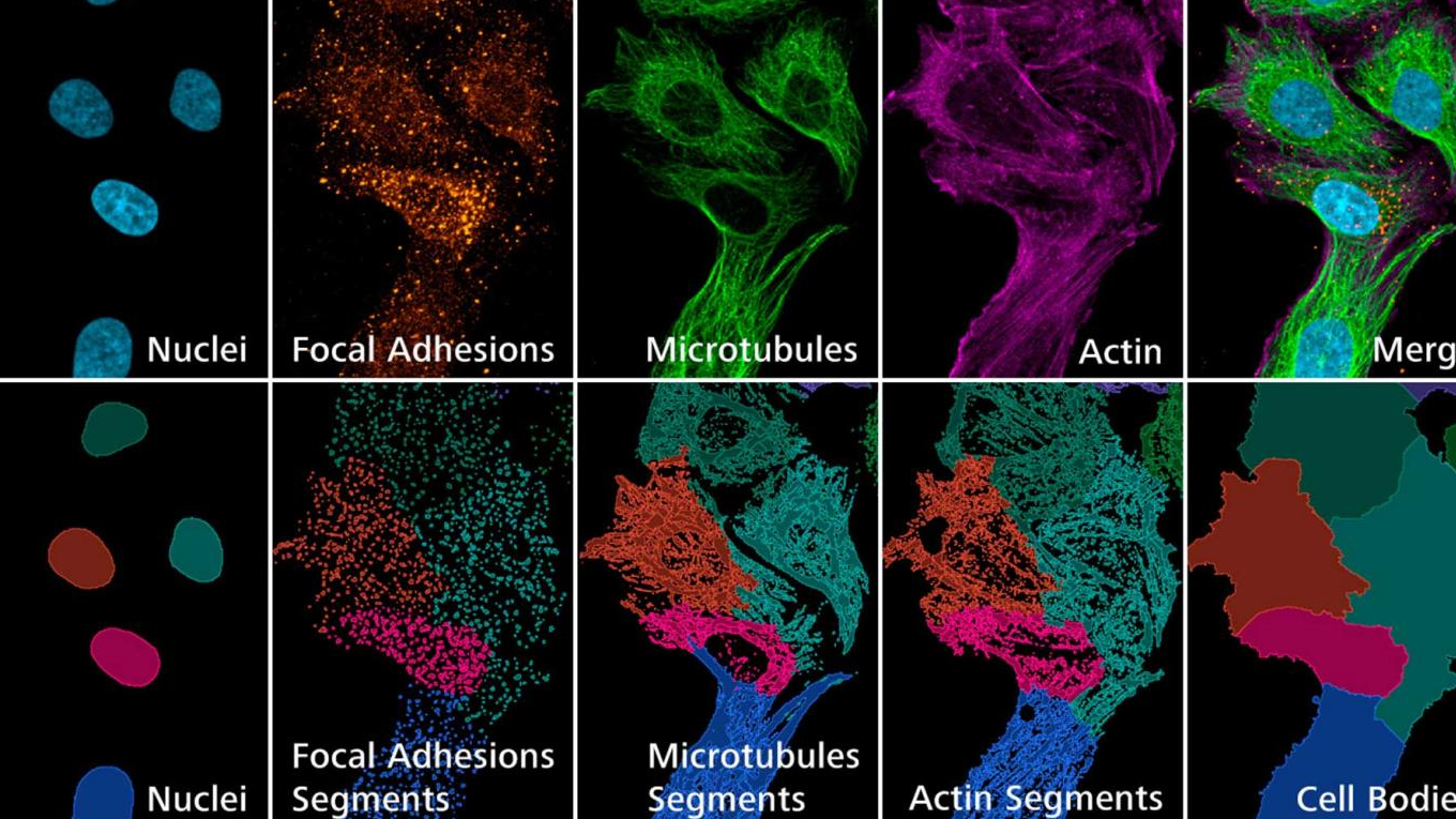
- Detect an ever-growing number of features in the cell, using automated image segmentation and analysis pipelines
- Allocate child features to the correct parent cell
- Use advanced data analysis tools for further statistical analysis.
ZEISS arivis Software
The multi-dimensional image analysis platform from ZEISS. Its flexible concept allows any researcher to put together image analysis workflows to answer creative questions for your research. Train AI models on the cloud (or locally) for image segmentation, set up end-to-end automated pipelines and scale up your analysis and store your data in one place.