Iodine-Contrast MicroCT Imaging for Mouse Embryo Development from Early Post-implantation to Early Postnatal Stages


Baylor College of Medicine
Iodine-Contrast MicroCT Imaging for Mouse Embryo Development from Early Post-implantation to Early Postnatal Stages
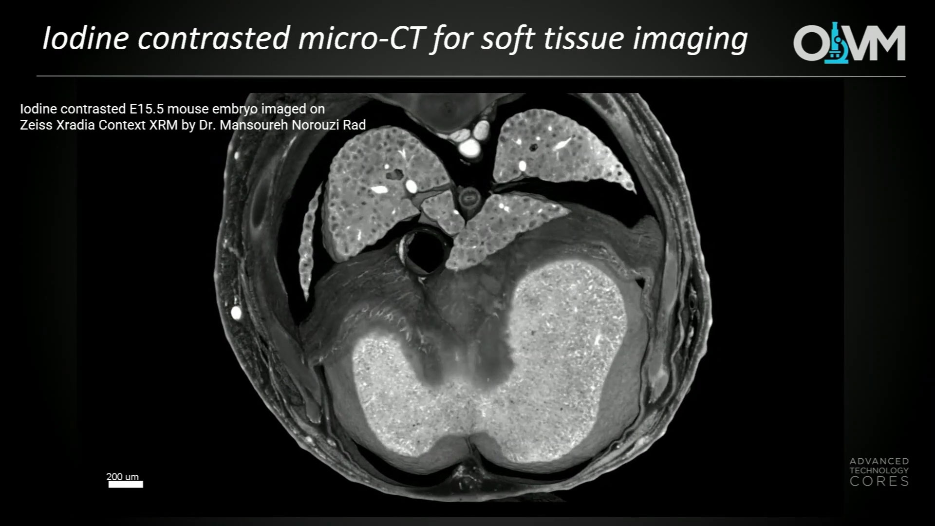
Mouse models have long been a crucial tool for studying human disease, However, traditional methods of studying mouse embryos and phenotypes have been limited in their resolution and scope. The use of iodine contrast microCT has revolutionized our understanding of early development and embryonic abnormalities in mouse models. MicroCT imaging allows for non-destructive, 3D visualization of tissues and organs at high resolution, enabling researchers to study embryonic development and abnormalities with unprecedented detail.
As part of The International Mouse Phenotyping Consortium (IMPC), we have utilized iodine-contrast microCT to analyze mouse embryos and neonates from embryonic day 8.5 to postnatal day 3, while demonstrate that early-stage embryos can also be imaged without disturbance of extra embryonic tissues. This method allows for comprehensive analysis of embryo development, providing valuable insights into embryonic lethality in knockout mouse lines. Additionally, these techniques are cost-effective, easy to learn, and time- efficient, making them ideal for high-throughput analysis.
Key Learnings:
- Comprehensive analysis of embryo development can be achieved using X-ray microCT
- Established methods for sample preparation, including iodone contrast, make this approach ideal for high-throughput analysis.
- The non-destructive assessment of iodine contrasted specimens has revolutionized current understanding of early development and embryonic abnormalities in mouse models.