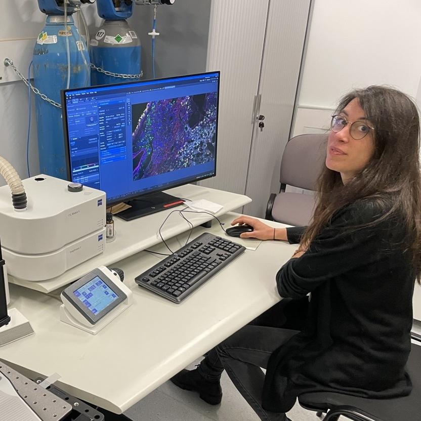
Spectral imaging with the ZEISS LSM 980 laser scanning confocal enables more complex studies of cell-cell interactions and locations in immunology research.
The immune system is a complex network of cells, tissues, and organs that works together to form multitier responses to foreign pathogens and abnormal cells. Immune cells frequently interact through close cell-to-cell contact. Spatial biology imaging studies using fluorescent markers to identify different immune cell types can provide insights into important cell-cell interactions and diverse microenvironments. However, experimental designs have typically been limited to three to four fluorescent labels based on the imaging set-up capabilities. Additionally, researchers working with tissues are often challenged with autofluorescence, which can contaminate fluorescent measurements resulting in misinterpretation of imaging data.
Spectral microscopy is one solution not only for imaging higher numbers of fluorophores but also for removing autofluorescence signals. Dr. Sandra Ormenese, Alexandre Hego, and Gaëtan Lefevre work at the GIGA Cell Imaging & Flow Cytometry Platform, a core imaging facility supporting researchers in cancer, neuroscience, cardiology, and immunology at the University of Liège, Belgium. They have implemented spectral microscopy with the ZEISS LSM 980 laser scanning confocal to enable researchers working with lung immunity to expand their research in spatial biology studies with up to eight fluorescent markers.
We started spectral imaging with LSM 980 for a spatial biology project studying allergic asthma in lung tissue with 6 fluorescent markers. Now more and more researchers ask us to design similar experiments. We have a recent success with 8 fluorescent markers for a project examining lung repair after injury.
Six labels were used to identify immune cells in lung tissue: DAPI (blue), IL-5 (red), CD3 (green), CD8 (white), CD11b (purple), and CD11c (yellow). Spectral imaging was performed with the ZEISS LSM 980 laser scanning confocal.
Six labels were used to identify immune cells in lung tissue: DAPI (blue), IL-5 (red), CD3 (green), CD8 (white), CD11b (purple), and CD11c (yellow). Spectral imaging was performed with the ZEISS LSM 980 laser scanning confocal.
6-COLOR SPECTRAL IMAGING
Deciphering Subpopulations of Immune Cells in the Lung
The hygiene hypothesis states that exposure to certain microorganisms during childhood modulates the development of allergic diseases such as asthma. In this context, researchers from Drs. Bénédicte Machiels and Laurent Gillet's Immunology-Vaccinology Laboratory were interested to study the impact of respiratory infection by a gammaherpesvirus on alveolar macrophages and innate lymphoid cells type 2 (ILC2) in the lung.
Spectral microscopy with LSM 980 was employed to image lung tissue that had been labeled with more markers than typically used for spatial biology studies to identify a more diverse cell population set. Their approach expanded the experimental design to six markers including IL5, CD3, CD68, CD11b, CD11c, and DNA.
While identifying ILC2s and deciphering the heterogeneity of myeloid cells in the lung is a major challenge, spectral imaging highlighted the existence of a key functional dialogue between ILC2s and alveolar macrophages that is responsible for variations in individual susceptibility in the development of allergic asthma.
Lungs are an organ with significant autofluorescence. Pre-treatment along with the LSM 980 performance made these studies possible. Spectral imaging was crucial in revealing cellular interactions within the pulmonary tissue, underscoring the utility of such tools in immunological research.
Eight labels are used to identify various macrophage population niches: pSPC (AT2) (yellow), MHC2 (magenta), CD68 (cyan), podoplanin (AT1) (white), CD31 (red), Ki67 (green), Sytox Blue (nuclei) (blue). The 8th "label" is the autofluorescence channel which is not shown in the image. Spectral imaging was performed with the ZEISS LSM 980 laser scanning confocal.
Eight labels are used to identify various macrophage population niches: pSPC (AT2) (yellow), MHC2 (magenta), CD68 (cyan), podoplanin (AT1) (white), CD31 (red), Ki67 (green), Sytox Blue (nuclei) (blue). The 8th "label" is the autofluorescence channel which is not shown in the image. Spectral imaging was performed with the ZEISS LSM 980 laser scanning confocal.
8-COLOR SPECTRAL IMAGING
Identification of Macrophage Niches in Wounded Lungs
The lung is constantly exposed to airborne pathogens and particles that can cause alveolar damage. One area of research in the Immunophysiology Laboratory headed by Dr. Thomas Mariachal is to understand how myeloid cells develop and how these cells interact with structural cells to regulate tissue repair responses. Macrophages can adopt a broad range of distinct functional identities that depend on their differentiation trajectory, the diseased tissue microenvironment, the phase of inflammation, and their activation state.
Cecilia Ruscitti, a PhD studentin the Immunophysiology Laboratory, is heading a project to use an in vivo model of influenza A virus-triggered injury to investigate the spatiotemporal trajectory and function of macrophage populations.
With the LSM 980 spectral technology, an eight-label panel was designed to identify various macrophage population niches as well as the distribution of these cells throughout injured lung tissue and their interactions with structural cells. One particularly interesting observation was the atypical macrophages' perilesional distribution and their colocalization with regenerating epithelial cells. This suggested a role for them in lung regeneration, a hypothesis that they were able to confirm in further functional experiments.
The characterization of macrophage spatial distribution within the wounded lung was challenging. Multispectral technology enabled the simultaneous identification of various structural and immune cell markers, leading to a deep, image-based characterization.