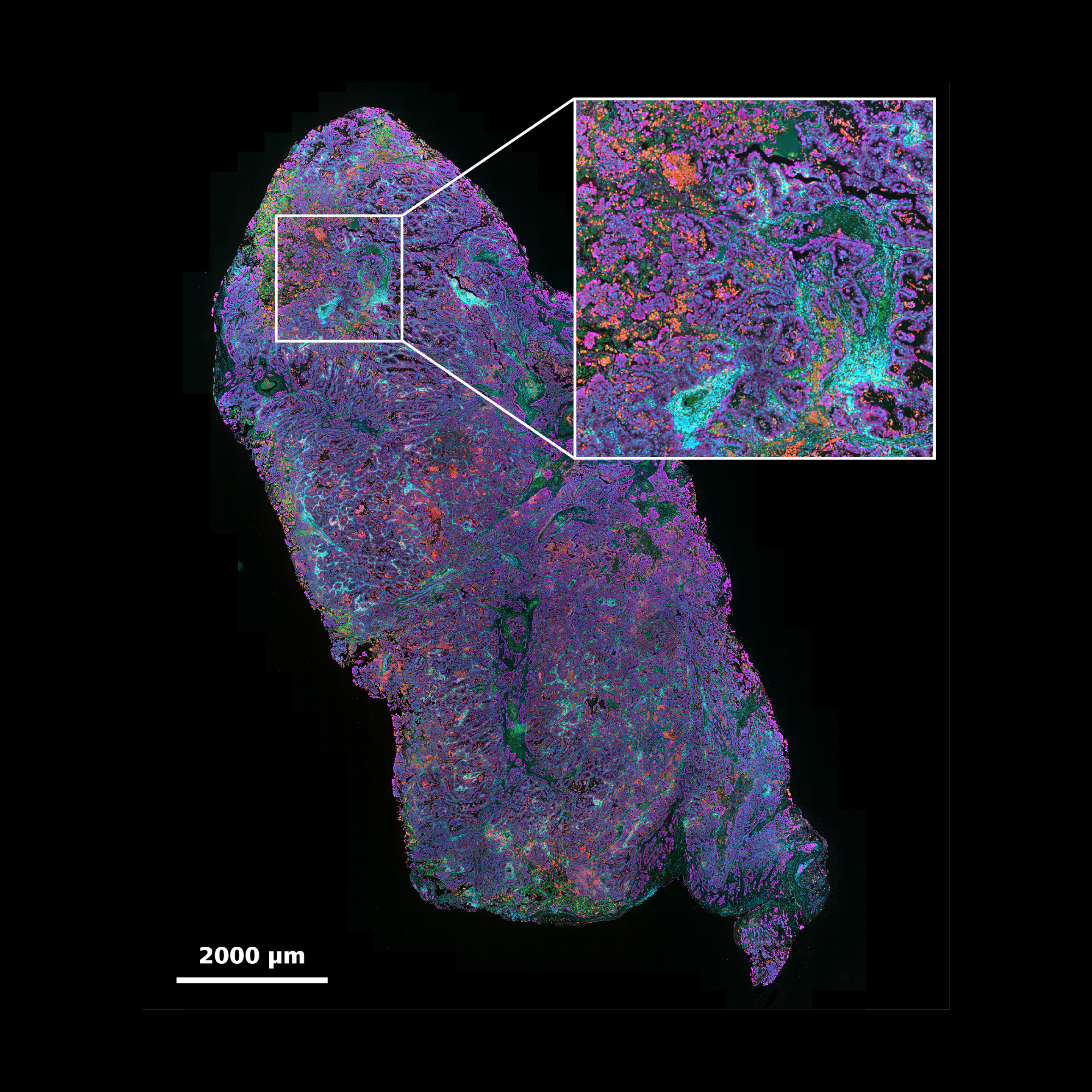
Visualize and analyze the complete tumor microenvironment from single-cell interactions to tissue-wide spatial patterns with multi-modal imaging solutions.
Accelerate Drug Discovery
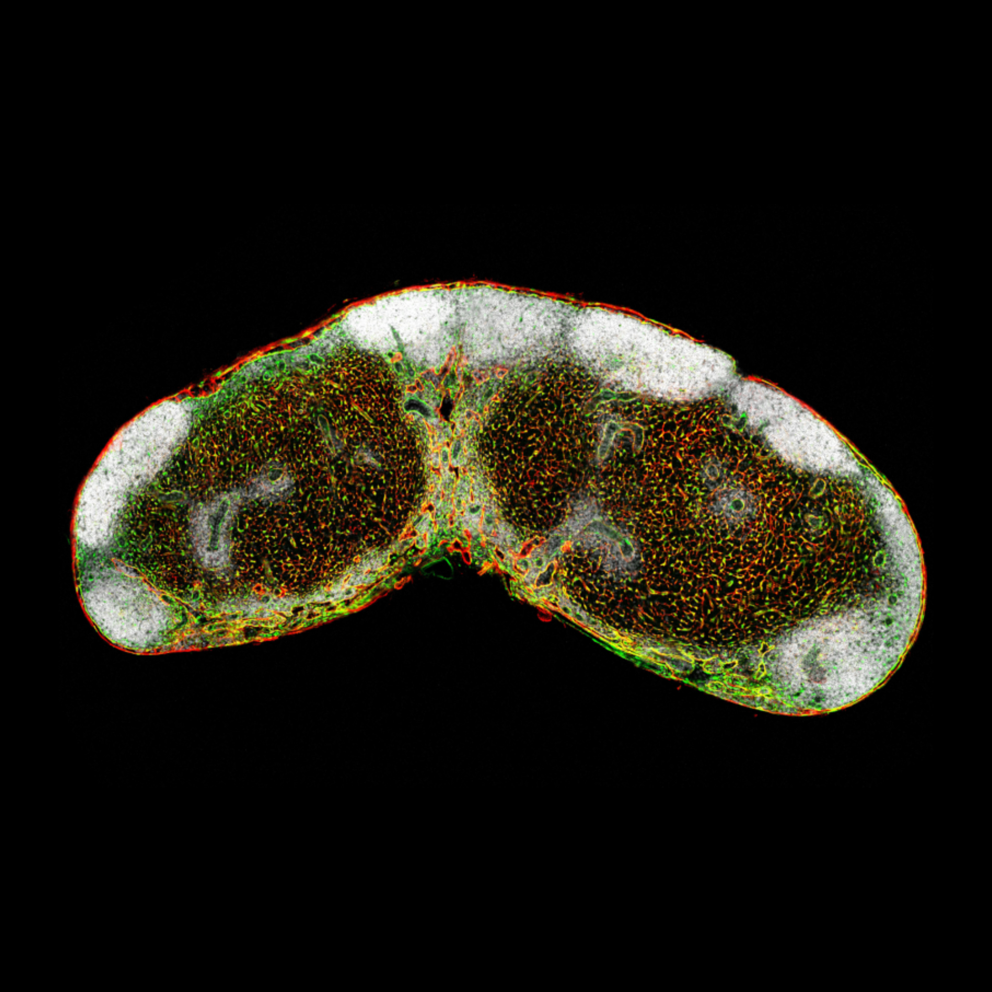
Screen drug candidates efficiently using 3D organoid models with automated imaging and analysis designed for high-throughput therapeutic evaluation.
Illuminate Every Biomarker
Uncover critical protein signatures and rare cell populations with advanced multiplex imaging and unprecedented sensitivity across multiple fluorescent channels.
Pushing the Boundaries in Cancer ResearchHarness cutting-edge microscopy techniques to explore the intricate mechanisms underlying tumor biology and progression. New innovative approaches enhance understanding of cancer cell behavior, microenvironment interactions, and therapeutic responses, paving the way for breakthroughs in personalized medicine and targeted therapies.
Scroll animation items
Are you ready for your next breakthrough?
Discover the power of high-resolution imaging and multiplex analysis
Reveal cancer's true architecture
Capture every detail in complex 3D organoid models with intelligent automation that accelerates discovery.
Maximize your multiplexing capabilities with superior spectral resolution that enables simultaneous visualization of numerous markers within a single specimen.
A single patient sample will create multiple slides – up to 40 slides depending on a test – so efficient digitization of slides is critical.
The integration of the ZEISS digital slide scanner into the Epic Sciences imaging platform has enabled us to improve our throughput and efficiency.
Recommended Products for Cancer Research
High-performance Slide Scanner for Fluorescence, Brightfield and Polarization
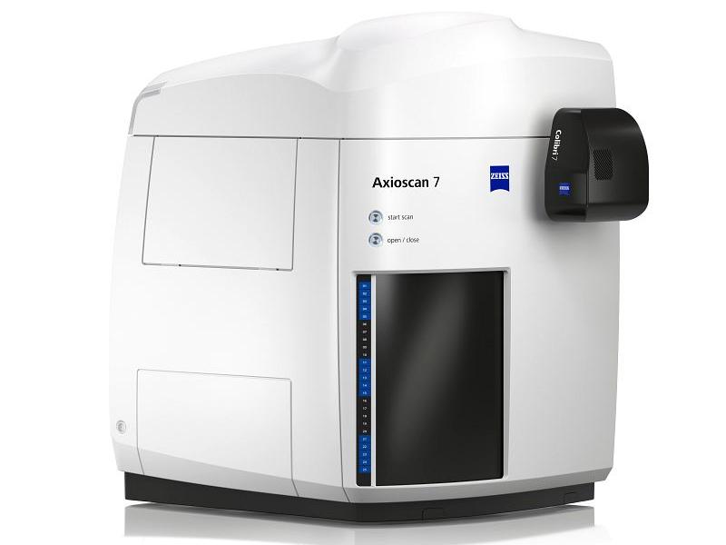
ZEISS Axioscan 7
ZEISS Axioscan 7 combines qualities that you would not expect to get in a slide scanner: high speed digitization and outstanding image quality plus an unrivaled variety of imaging modes are all available in a fully automated and easy to operate system.
Multiple imaging modalities, including brightfield, fluorescence, and phase contrast imaging.
Can scan up to 1,000 slides in a single run, significantly enhancing throughput for large-scale studies.
Sophisticated filter concept for demanding fluorescence imaging.
Adaptable Automation for Advanced Workflows
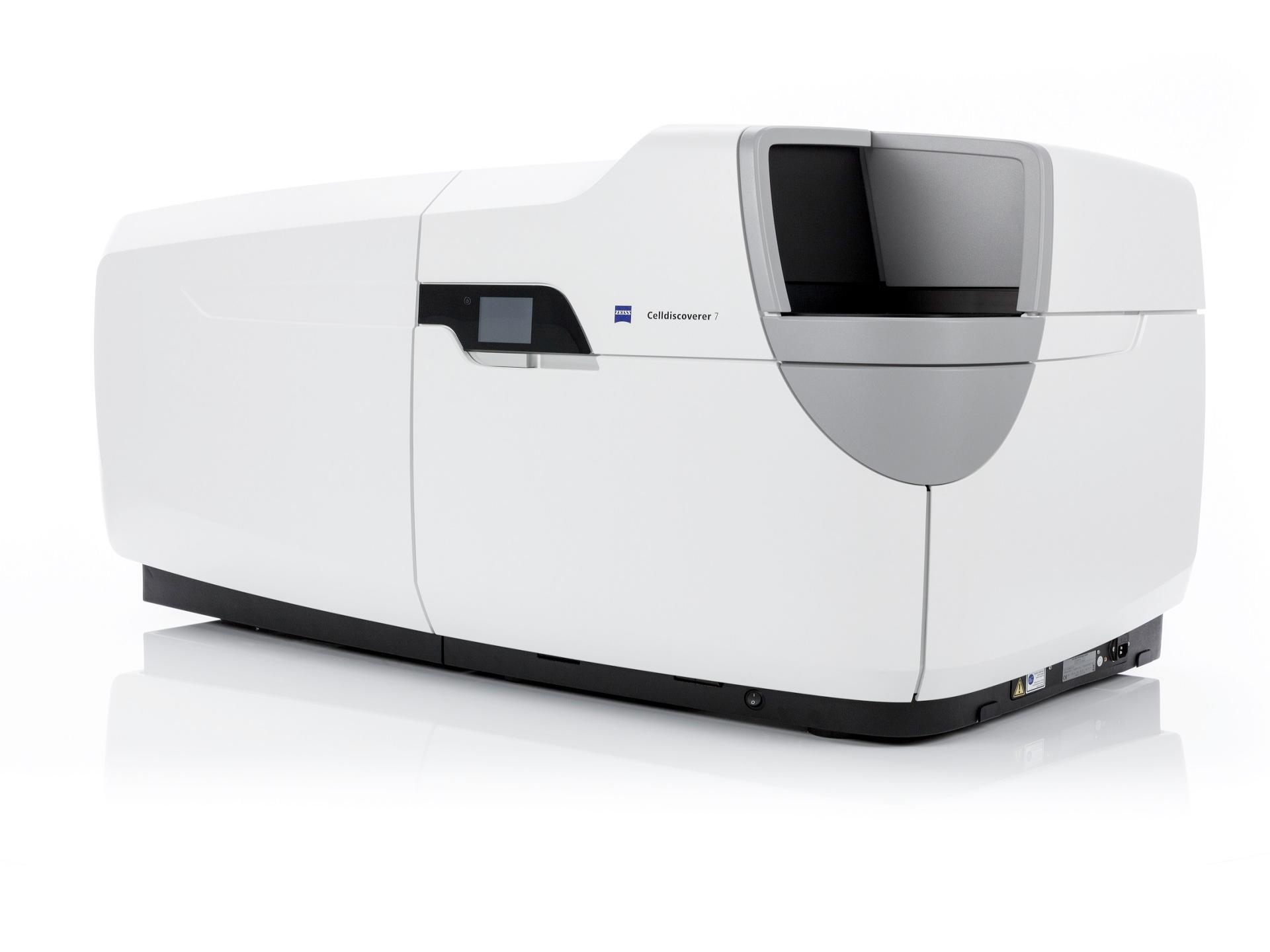
ZEISS Celldiscoverer 7
ZEISS Celldiscoverer 7 is your research companion for collecting statistically relevant data, giving you easy access to high-quality imaging, adaptability to demanding experiments, and stable long-term operation.
Full support of auto-immersion objectives significantly improves Guided Acquisition workflows.
Reliable in multi-user environments.
Top-Class Multimodal Imaging Combined in One Confocal System
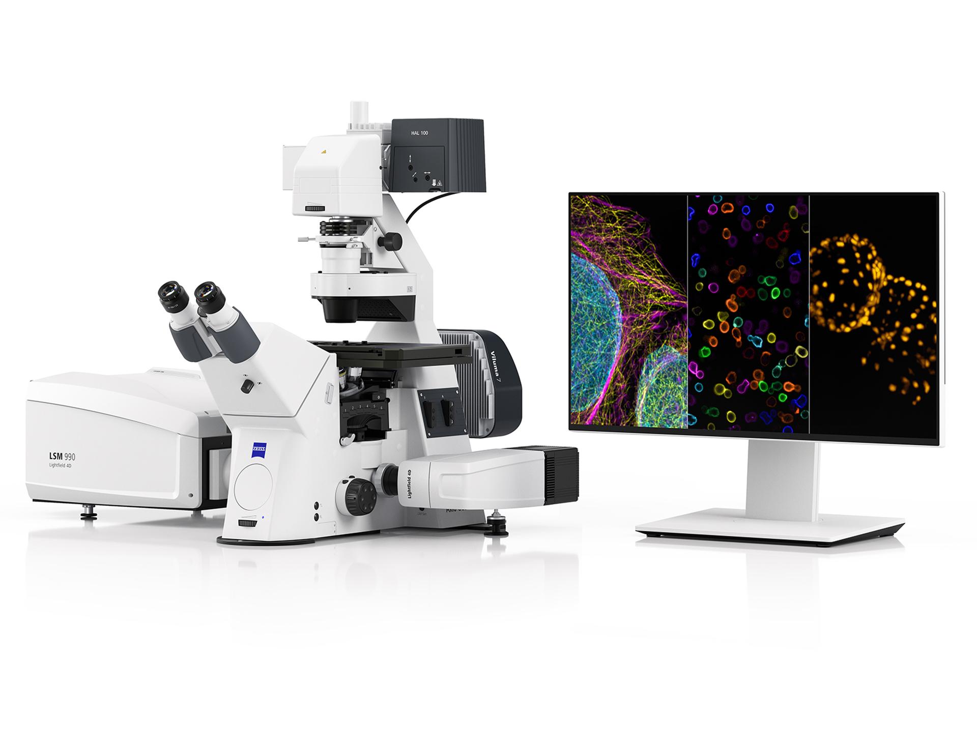
ZEISS LSM 990
ZEISS LSM 990 combines an unprecedented range of imaging options to help you explore new research dimensions. Delve into your biological research with 90 nm super-resolution, high-speed volumetric imaging, or the separation of 10 fluorescent labels simultaneously.
Capture entire model organisms and tissue sections
Acquire super-resolution images as fast and gentle as widefield images
Go from a large-field overview to the super-resolution details
Long-Term Volumetric Imaging of Living Cells
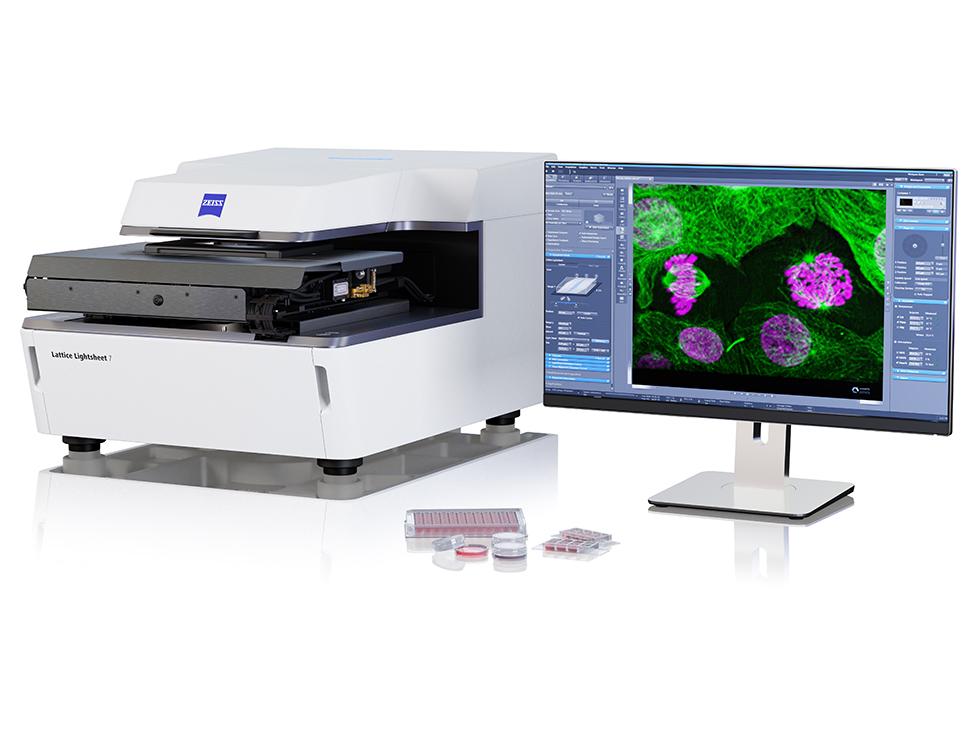
Lattice Lightsheet 7
ZEISS Lattice Lightsheet 7 makes light sheet fluorescence microscopy available for live cell imaging at subcellular resolution – while also allowing you to use your standard sample carriers.
Examine living specimens directly on your standard sample carriers
Watch the subcellular dynamics of life over hours and even days
Reveal three-dimensional details in their true proportions
Your All-in-One Cell Imaging System
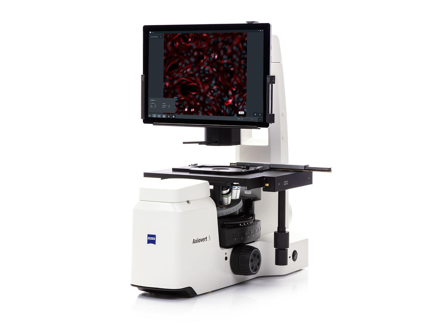
ZEISS Axiovert 5 digital
Axiovert 5 digital brings AI into your cell lab to ease your daily work. It will make your processes more efficient and your results more reproducible.
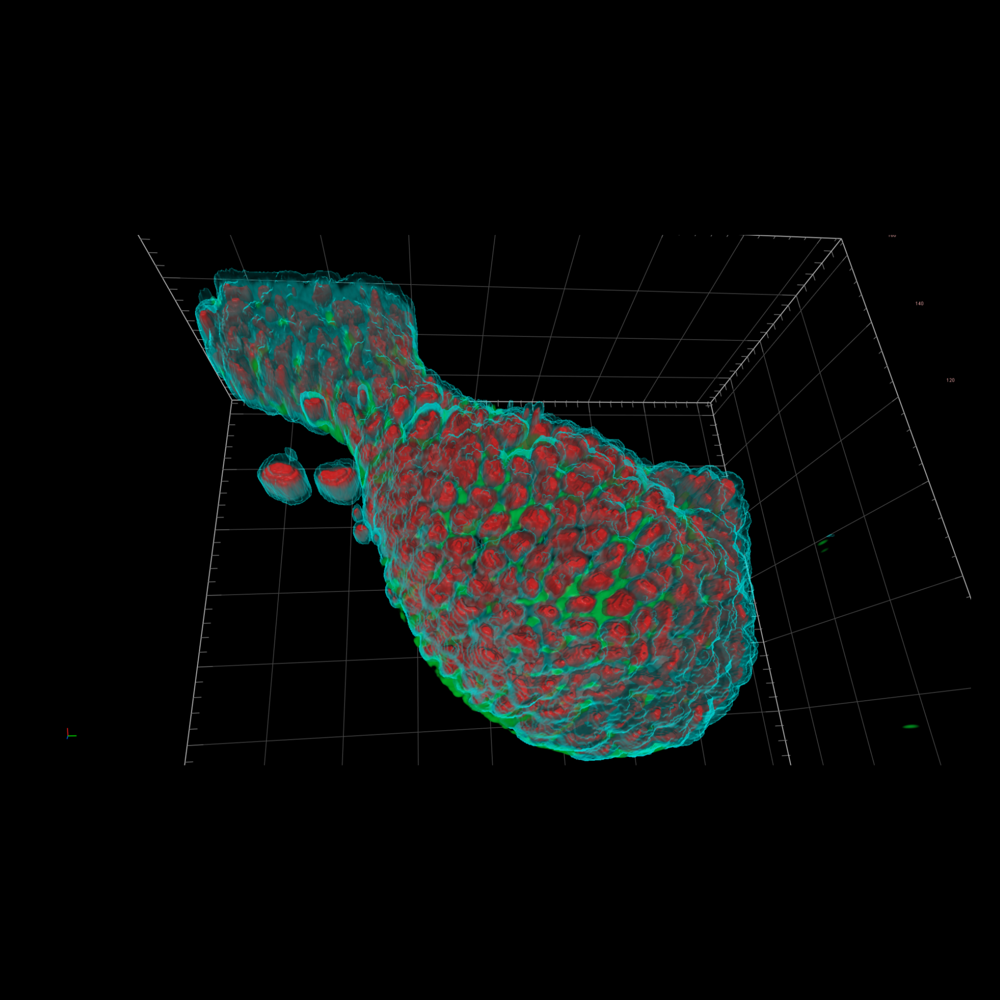
The ZEISS Celldiscoverer 7 combined with arivis Pro software provides an automated solution for high-content organoid analysis. The system can automatically detect and focus on organoids, segment their outer layer and lumen, and measure key parameters like volume, shape, and cell numbers. For more complex analyses, the system can track marker expression at the single-cell level and process hundreds of organoids in multi-well plates.
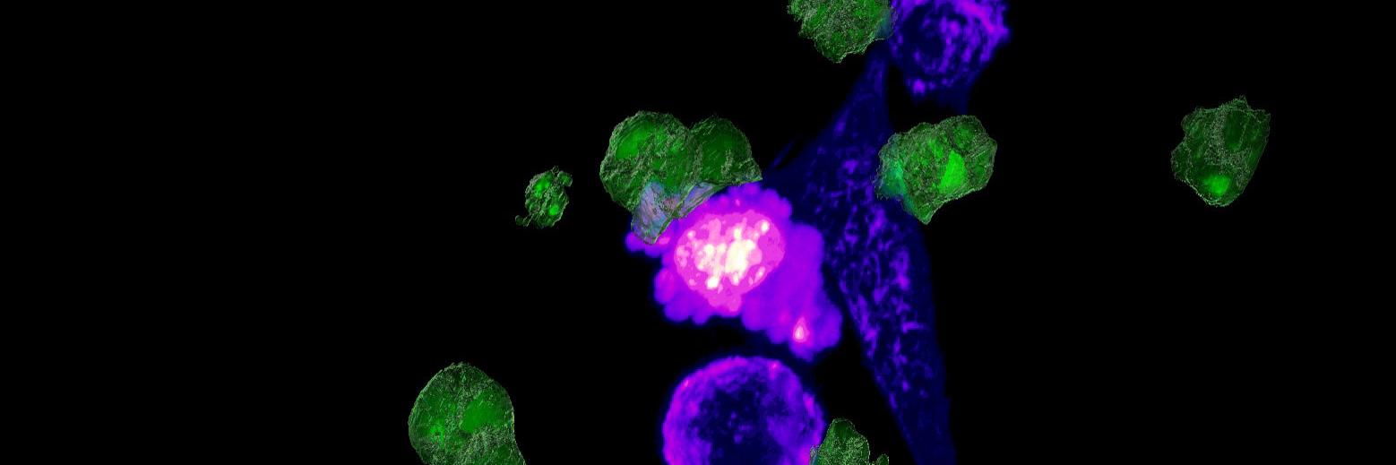
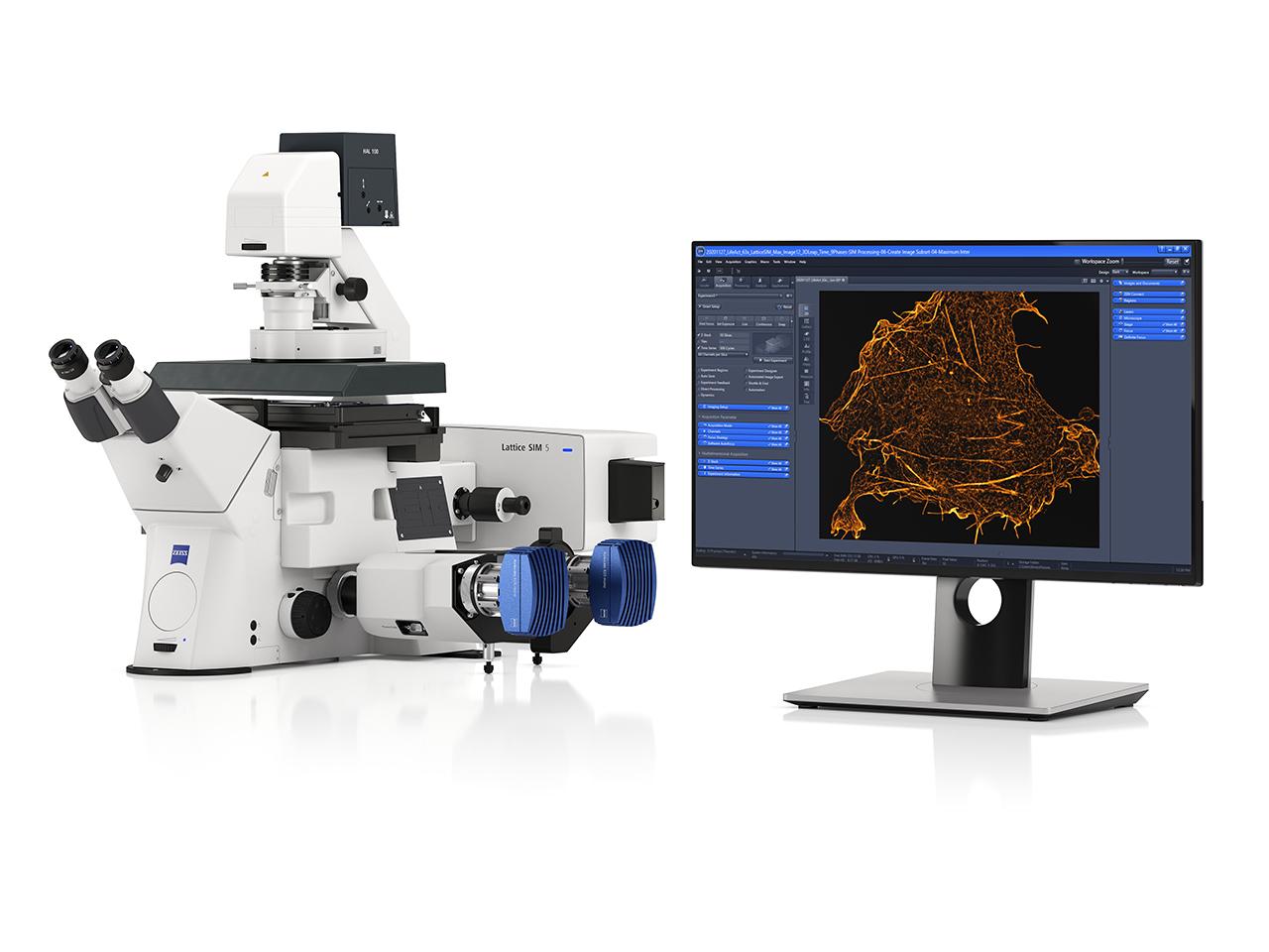
The ZEISS LSM 980 with Airyscan 2 offers exceptional sensitivity and spectral flexibility for multi-channel imaging of the tumor microenvironment. The system provides super-resolution capabilities down to 120 nm laterally, while the new Multiplex mode enables faster acquisition for capturing dynamic interactions. For even higher resolution of subcellular structures, the new Lattice SIM 5 can achieve 60 nm resolution with up to 4 channels.
The ZEISS Axioscan 7 is ideal for high-throughput digital pathology and biomarker screening, capable of scanning up to 100 slides unattended. The system supports brightfield, fluorescence and polarization imaging modes, with automated focusing and scan profile creation through ZEN Slidescan software. Data management and sharing is streamlined through ZEN Data Storage and Explorer.
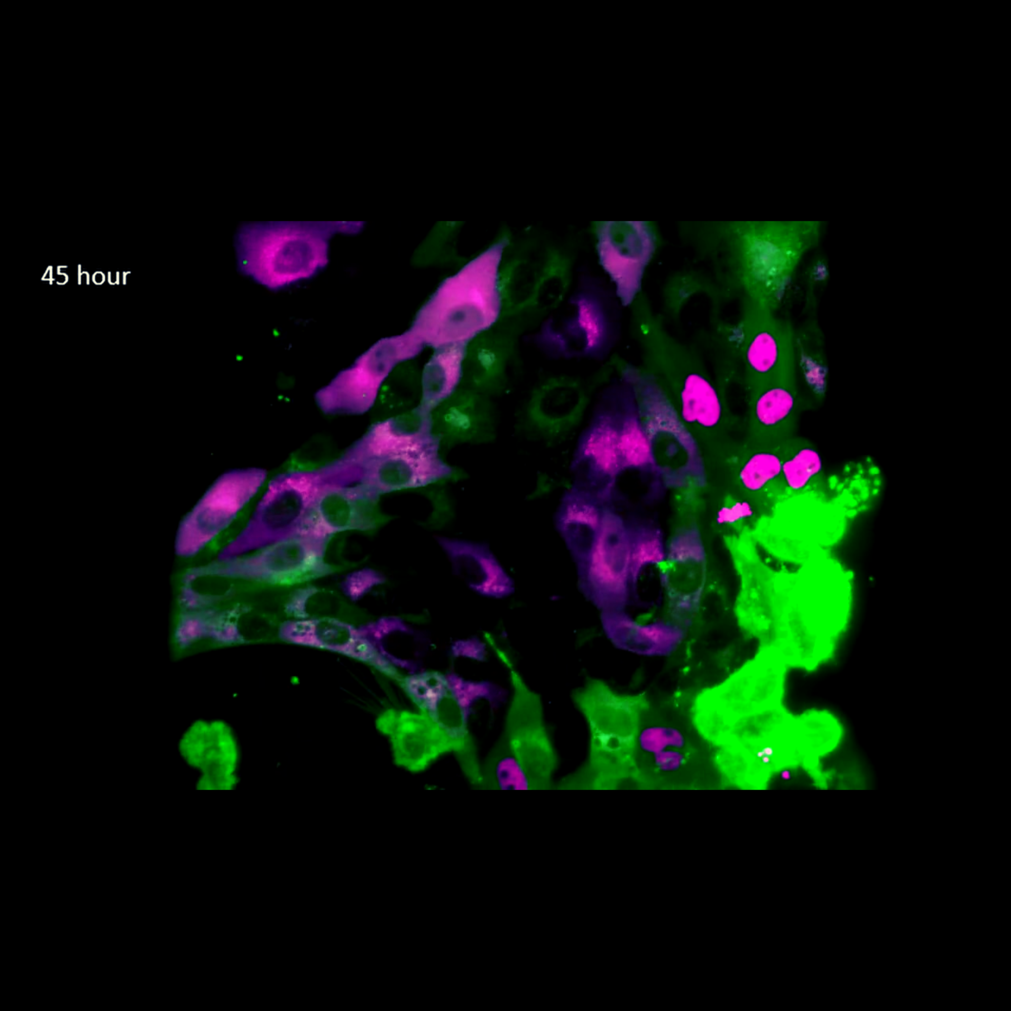
The ZEISS Lattice Lightsheet 7 is specifically designed for gentle, long-term volumetric imaging of living samples. The system uses an ultra-thin light sheet that minimizes phototoxicity while providing extremely fast acquisition speeds. Built-in environmental controls maintain optimal conditions for multi-day imaging of cell migration and invasion processes.
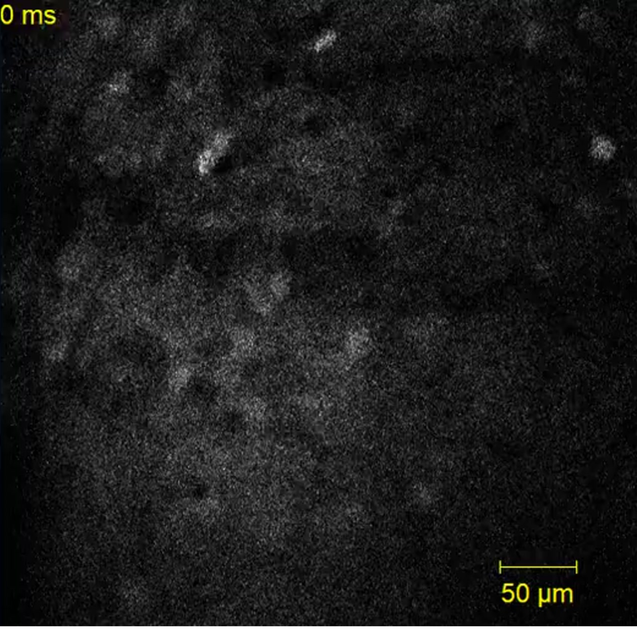
The new ZEISS Lattice SIM 3 combines fast imaging speeds (up to 255 fps) with 120 nm spatial resolution, making it ideal for capturing rapid immune cell-tumor cell interactions. For longer term studies, the Celldiscoverer 7 with confocal module provides automated environmental control and multi-position acquisition. Both systems can be combined with incubation and environmental control for maintaining physiological conditions during extended imaging sessions.
Comprehensive Solutions and Capabilities
Discover Our Application Hub
Explore applications to discover tailored solutions for your unique laboratory needs and elevate your research capabilities.