fluorescence human-kidney Celldiscoverer-7
Sample courtesy of Sanofi-Aventis Deutschland GmbH; R&D IDD / in vitro Biology, Frankfurt, Germany
Use Case

Cell Culture Research

Explore the frontier of cell culture technology with advanced microscopy solutions. Enhance your laboratory's capabilities in biomedical research, biotechnology, and more, with state-of-the-art instruments designed for precision and efficiency.


Empowering Research with Advanced Microscopy

Enhancing Cell Culture Observations

  • cell culture
  • cell culture
  • Microglia uptake of synaptic proteins within the zebrafish hindbrain at 28 days post fertilization (dpf).
    Microglia uptake of synaptic proteins within the zebrafish hindbrain at 28 days post fertilization (dpf).
    Sample courtesy of Michael W. Davidson, The Florida State University
    Sample courtesy of Michael W. Davidson, The Florida State University
  • Cell Culture
    Cell Culture
    Sample courtesy of P. Denner, Core Research Facilities, German Center of Neurodegenerative Diseases (DZNE), Bonn, Germany.
    Sample courtesy of P. Denner, Core Research Facilities, German Center of Neurodegenerative Diseases (DZNE), Bonn, Germany.

Advanced Tools for Microscopic Analysis

In the dynamic field of cell culture research, microscopy not only provides a window into the microscopic world but also empowers scientists to explore the intricacies of cellular behavior in unprecedented detail. Since the inception of phase contrast microscopy, developed by Frits Zernike and rapidly implemented by ZEISS, visual inspection of living cells has become foundational in laboratories worldwide. Today, the discipline benefits from a suite of advanced microscopy techniques including phase contrast, fluorescence microscopy, and automated imaging systems.

These technologies facilitate detailed observations of cell morphology, growth patterns, and genetic expressions in real-time. For example, the ZEISS range of microscopes and imaging solutions supports daily tasks such as monitoring cell viability and confluency, crucial for maintaining the integrity of culture conditions. Fluorescence microscopy, enhanced by LEDs to minimize phototoxic effects, allows researchers to label and visualize molecular components within cells, providing insights into cellular functions and processes.

Moreover, automated systems like ZEISS Celldiscoverer 7 streamline workflows, reducing manual intervention and maintaining high standards of reproducibility and precision. This integration of sophisticated imaging technologies ensures that researchers can observe and analyze cellular phenomena with clarity and precision, driving forward innovations in drug development, regenerative medicine, and beyond. The ongoing advancements in microscope technology thus continue to play a pivotal role in the evolution of cell culture methodologies, paving the way for groundbreaking discoveries in life sciences.

Complete your lab

with our new solution bundles

You can enhance your Cell Culture Research with the following solutions

Microscopy & Cell Culture Research

Lattice Lightsheet 7

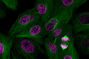
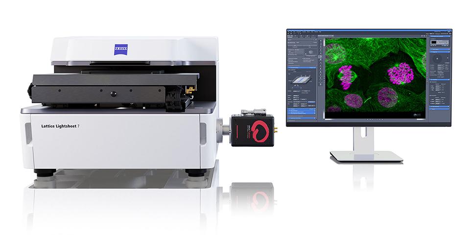
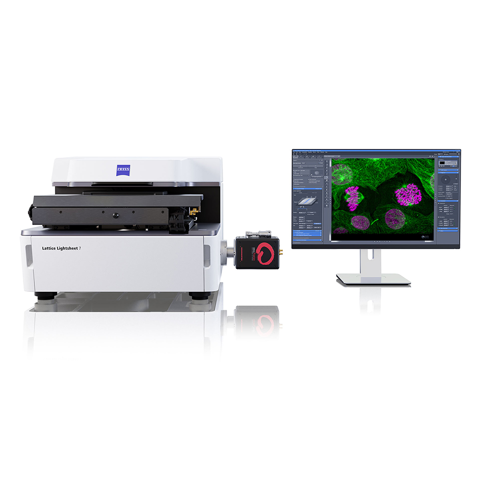
Long-Term Volumetric Imaging of Living Cells

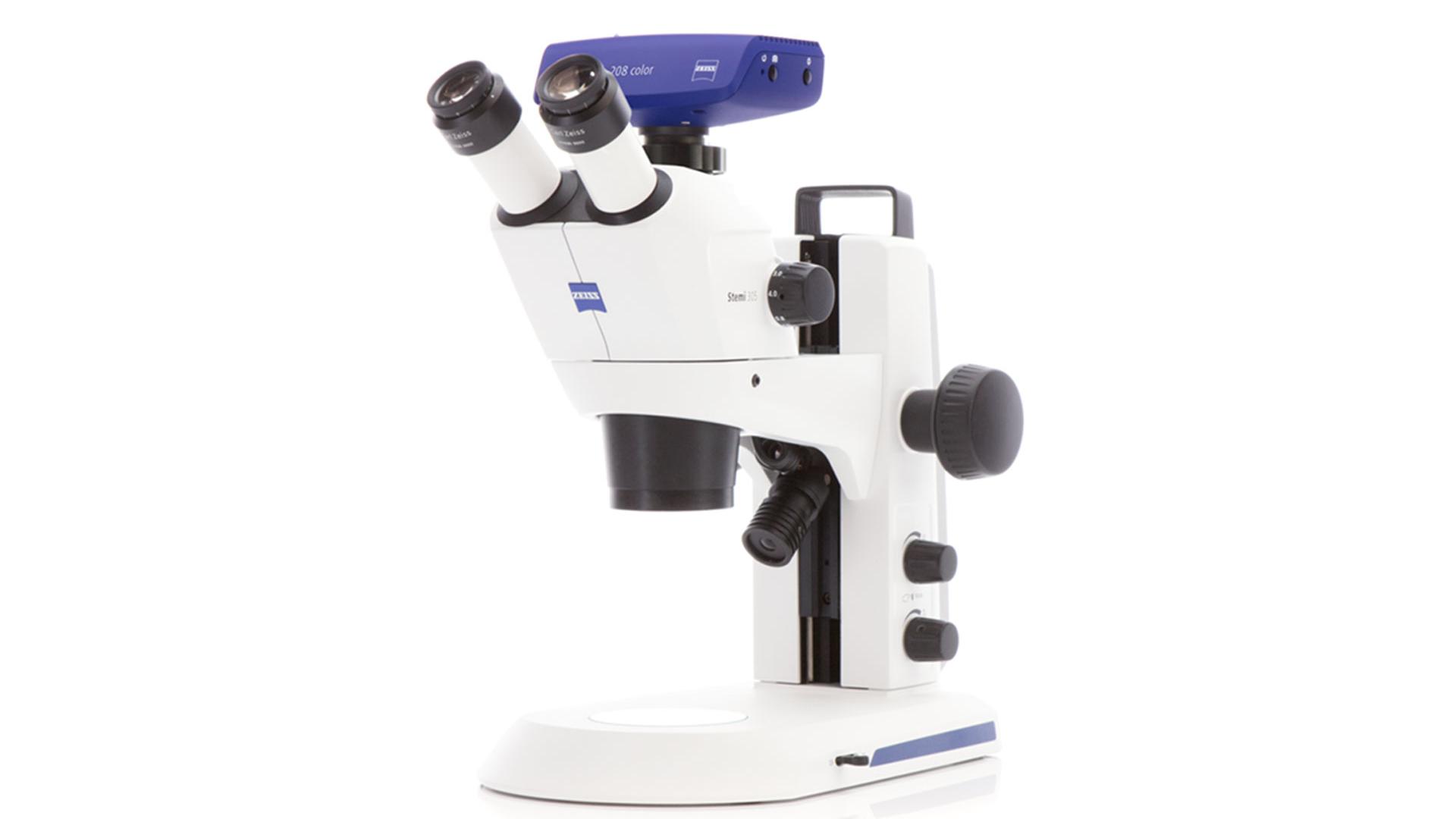
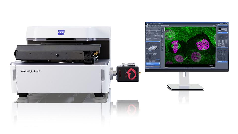
ZEISS microscopy technologies have been at the forefront of this revolution, offering tools that not only enhance the visual experience but also optimize research efficiency. From the pioneering phase contrast microscopy to the advanced Lattice Lightsheet 7, ZEISS provides a spectrum of microscopy solutions tailored for cell culture research. These systems are designed to handle live and fixed specimens with utmost care, reducing phototoxic effects and preserving the integrity of biological samples.
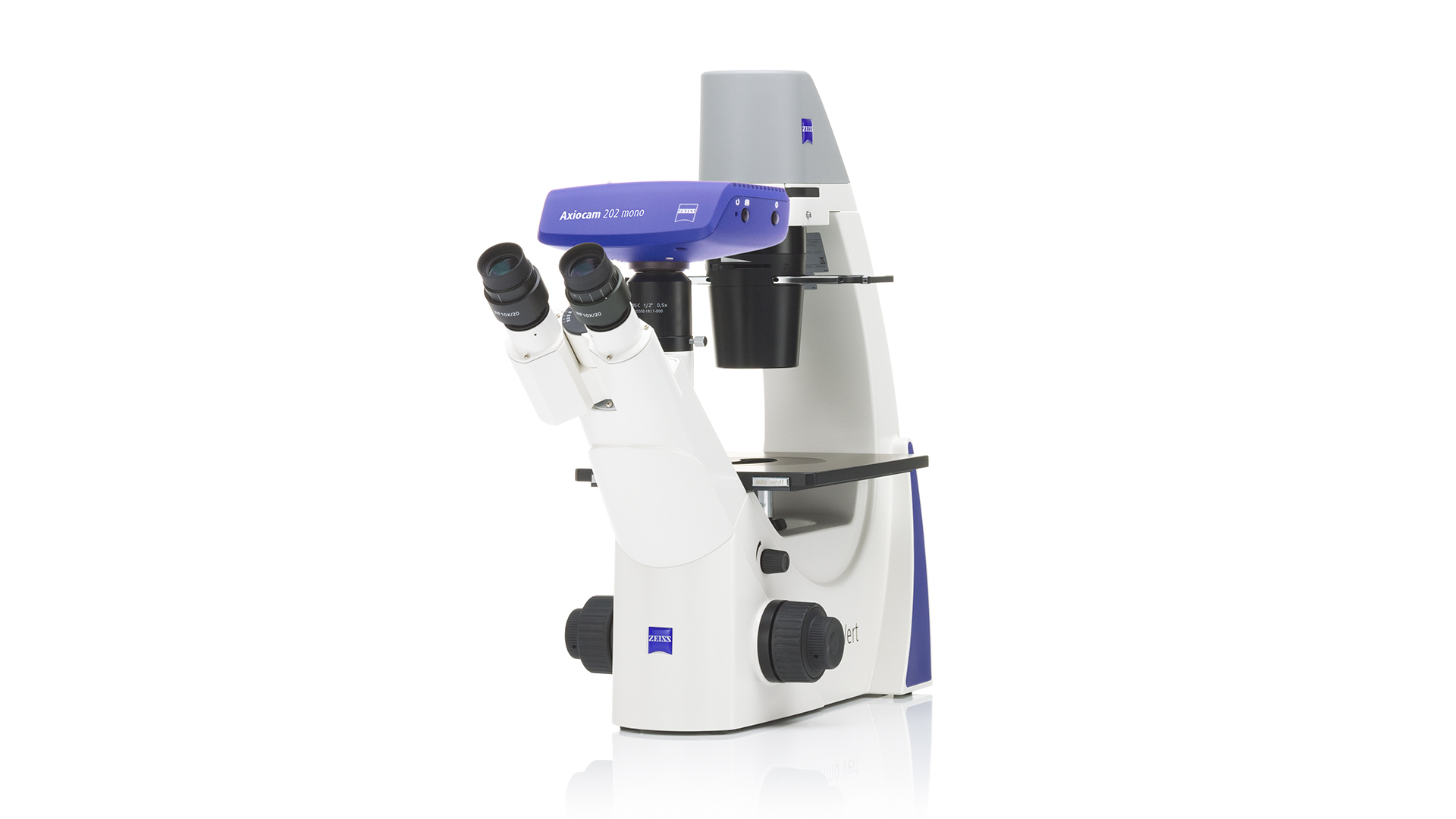
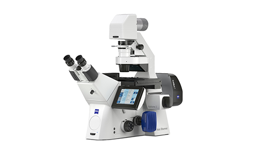
The integration of AI and automation in systems like the ZEISS Axio Observer and Celldiscoverer 7 transforms the daily routines of researchers into seamless workflows. These platforms offer flexibility, from simple widefield to sophisticated super-resolution imaging, catering to diverse research needs. Researchers benefit from features like automated sample detection, focus adjustments, and intuitive operation interfaces, which significantly cut down experimental setup times and enhance reproducibility.
 

Model Systems in Life Science Articles