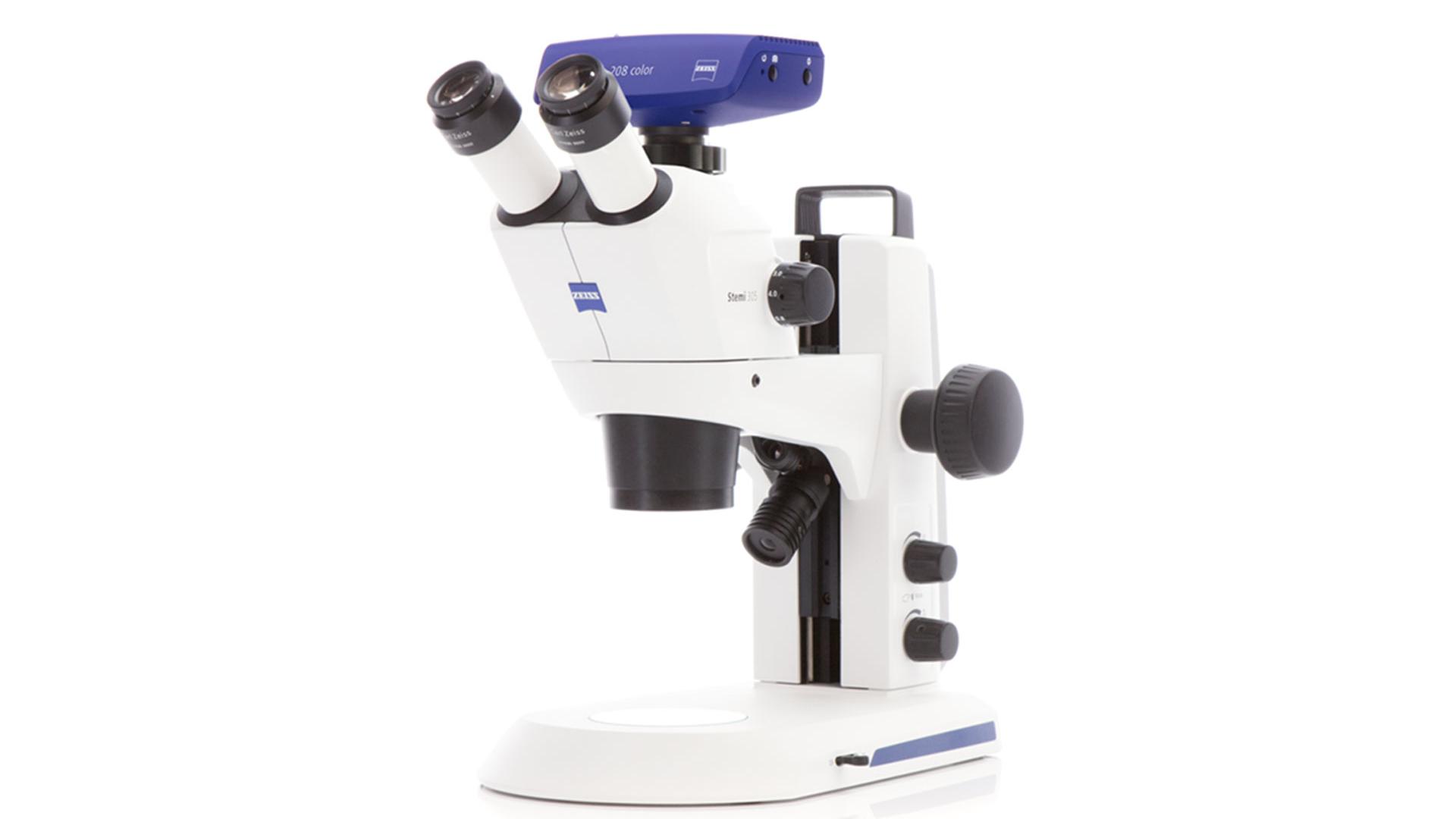
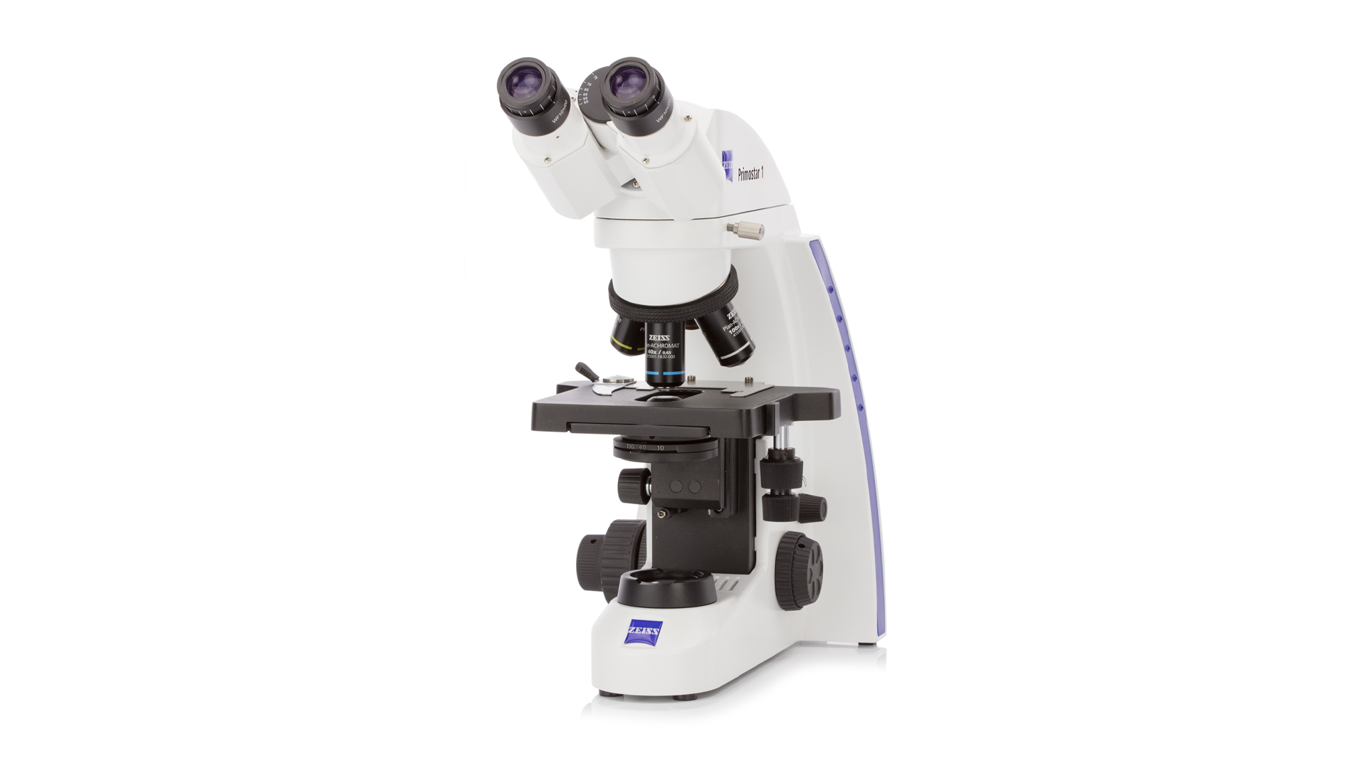
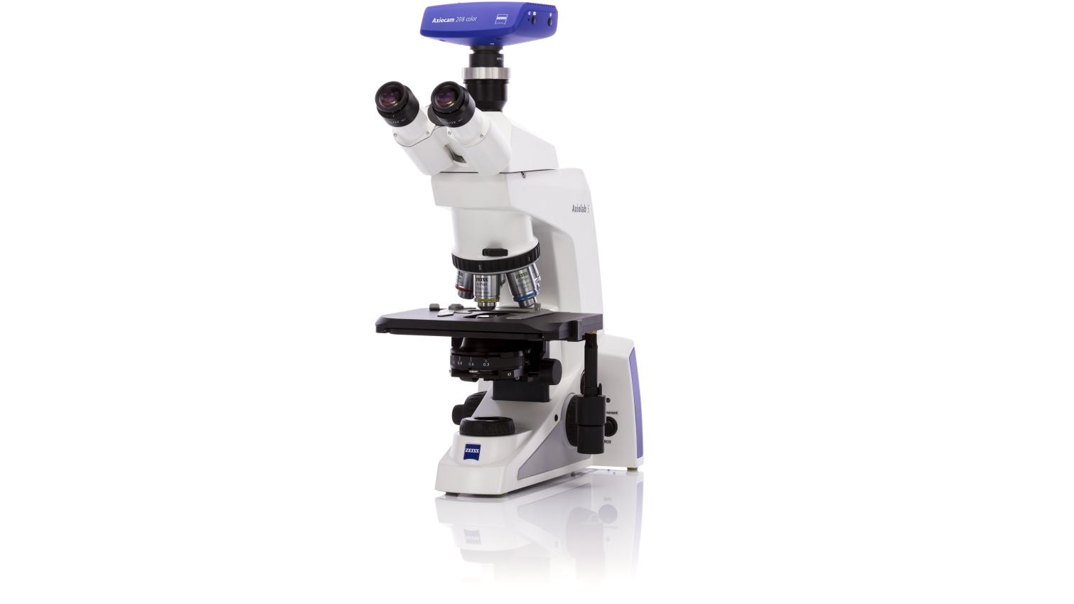
ZEISS microscopy applications revolutionize teaching and learning experiences in educational settings, from school systems to universities. With the shift towards remote learning, the demand for adaptable educational technologies has surged. ZEISS offers comprehensive solutions, including the Primostar 3 microscope equipped with integrated digital cameras and the Labscope app. This setup not only facilitates live streaming of microscopic sessions through platforms like Microsoft Teams or Zoom but also supports interactive learning by allowing students to engage with real-time images and videos, enhancing their understanding of complex scientific concepts.
Moreover, ZEISS Virtual Slide Box and ZEN Data Storage systems provide a unified platform where students can access and annotate microscopy slides remotely, making it easier for teachers to manage courses and maintain educational standards. The integration of these technologies ensures that students continue to receive a quality education, focusing on the detailed examination of samples which is crucial in life sciences.
Through ZEISS's digital tools, educators can keep students engaged and motivated, providing a near-real classroom experience that promotes active learning and continuous interaction. These solutions support not just the continuity of education but also its enhancement, by making complex scientific content more accessible and understandable. This holistic approach not only meets current educational needs but also sets a new standard for future learning environments in the sciences.