

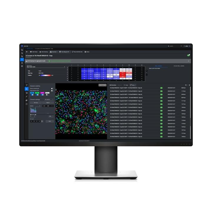
AI-Driven Image Analysis for Organoid Research and Analysis
With the FDA’s Modernization Act 2.0, organoids are taking center stage in vital research applications. In life sciences, biotech and pharma labs scientists are eager to adopt AI-driven approaches that will enable complex analysis of organoid growth, volume and quantification in 3D. Our innovative algorithms support your organoid research. From visualization to automated analysis and subcellular segmentation, we’ve got you covered. AI models help take your organoid analysis to new levels.


3D visualization of an organoid (green), nuclei in the outer cell layer (red) and nuclei in the lumen (yellow).
Cell-level Organoid Growth Analysis
Single-Cells Segmentation Within Organoids in 3D to Quantify Cell Numbers and Marker Expression for Organoid Growth AnalysisAutomated analysis is important to quantify the effects of drugs on growth of organoids and differentiation of cells within organoids.
This solution can be used to segment organoids, their lumen, and nuclei within the organoids using a combination of AI tools and conventional image analysis steps in ZEISS arivis Pro. Using the nuclei masks, the cell bodies of the individual cells within the outer layer of the organoids are segmented.
Having segmented the organoids and the cells within, several readouts are provided such as organoid and lumen volume, roundness of the organoids, and the number of cells per organoid. Furthermore, marker expression can be analyzed on a single-cell level and the percentage of marker-positive cells is reported. This analysis can be performed for individual images or for hundreds of images acquired using multi-well plates. With ZEISS arivis Hub the 3D organoid analysis is scaled up to include many different organoids.


3D Visualization of an organoid (green) and lumen (magenta), for volumetric analysis, which is displayed in a stacked column chart.
Organoid Volume Quantification
Analyze the Volume of Organoids and Their Lumen to Quantify Growth and DifferentiationAutomated analysis of organoid growth and differentiation is important for toxicity assays, drug screening and quality control protocols.
This solution can be used to quantify the volume of organoids and their lumens if applicable. As a first step, the organoid area and the lumen area are segmented using a trained deep learning network (trained on ZEISS arivis Cloud). This segmentation is then applied to z-stack images of many organoids gathered from multi-well plates.
For each organoid, 3D objects are generated that are used to quantify the organoid and lumen volumes. Finally, the measurements are presented as tables and interactive plots making it easy to understand and interpret the results.
Based on total volume, organoid growth was assessed. Based on the ratio of lumen per organoid, the differentiation of each individual organoid was assessed.


Organoid Image Analysis with the Power of AI to Boost Your Research
Recorded WebinarIn this webinar, our expert Dr. Sandra Lemke showcases how organoid images can be analyzed in 2D and 3D using the power of AI without the need to code. We will demonstrate an end-to-end pipeline setup that ensures reproducible results, which help you monitor organoid growth and differentiation.


Related Products


-
1
* The images shown on this page represent research content. ZEISS explicitly excludes the possibility of making a diagnosis or recommending treatment for possibly affected patients on the basis of the information generated with an Axioscan 7 slide scanner.