Neuroscience, the study of the nervous system and brain, has been transformed by recent advances in microscopy technologies.
These technological developments allow researchers to observe neural activities and structures with unprecedented detail. Advanced imaging techniques like High-Throughput Electron Microscopy and Light Sheet Fluorescence Microscopy provide comprehensive insights into neural connectivity and functionality..
These methods facilitate the creation of detailed neural maps, aiding in the understanding of complex brain functions and disorders. Furthermore, innovations in live tissue imaging offer real-time observation of neural dynamics, critical for studying neurodegenerative diseases. Coupled with computational modeling, these imaging technologies not only enhance our understanding of brain architecture but also propel forward the development of targeted neurological therapies.
Such breakthroughs underscore the importance of microscopy in neuroscience, making it indispensable for the ongoing exploration of one of the most complex entities in science—the human brain.
")
")


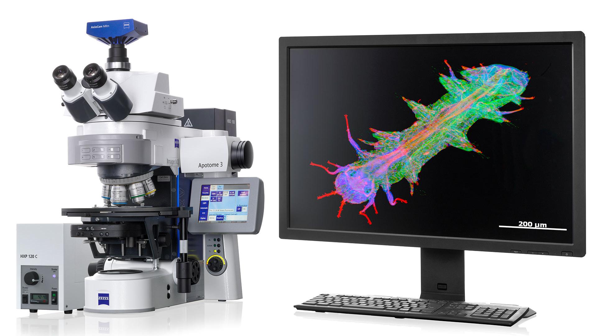
 Sample courtesy of Pr. Jim Trimmer Lab, University of California Davis and Neuromab")
 Sample courtesy of Pr. Jim Trimmer Lab, University of California Davis and Neuromab")
 Sample courtesy of Pr. Jim Trimmer Lab, University of California Davis and Neuromab")
 Sample courtesy of Pr. Jim Trimmer Lab, University of California Davis and Neuromab")