Multiscale Analysis – From Whole Plants to Subcellular Levels
Gain deeper insights into plant biology - visualize cellular structures, organelle dynamics, cell wall composition, and key processes such as growth, division, intracellular transport, and stress responses.
Spectral Flexibility for Complex Studies
Perform simultaneous imaging of multiple fluorescent markers while effectively separating autofluorescence in plant science samples.
Leverage automated image segmentation and AI-driven quantification for reproducible, high-throughput plant research.
Pioneering the Future of Plant BiologyFrom high-speed, high-resolution imaging and non-destructive 3D visualization, to real-time analysis, discover advanced microscopy techniques that capture both intricate and dynamic details of plant specimens. Explore complex structures and biological processes with precision, gaining deeper insights into photosynthesis, plant development, and responses to environmental stressors. Push the boundaries of discovery and innovation with cutting-edge imaging solutions.
Scroll animation items
Want to visualize the unseen world of plant biology?
Discover techniques to transform your research
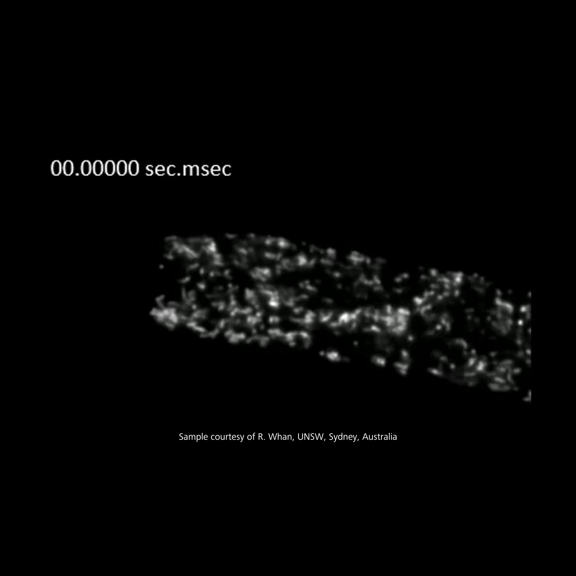
Long-Term Imaging of Living Cells
Break free from the limitations of traditional imaging and explore live plant cells as they truly are—in motion, in 3D, and in real time.
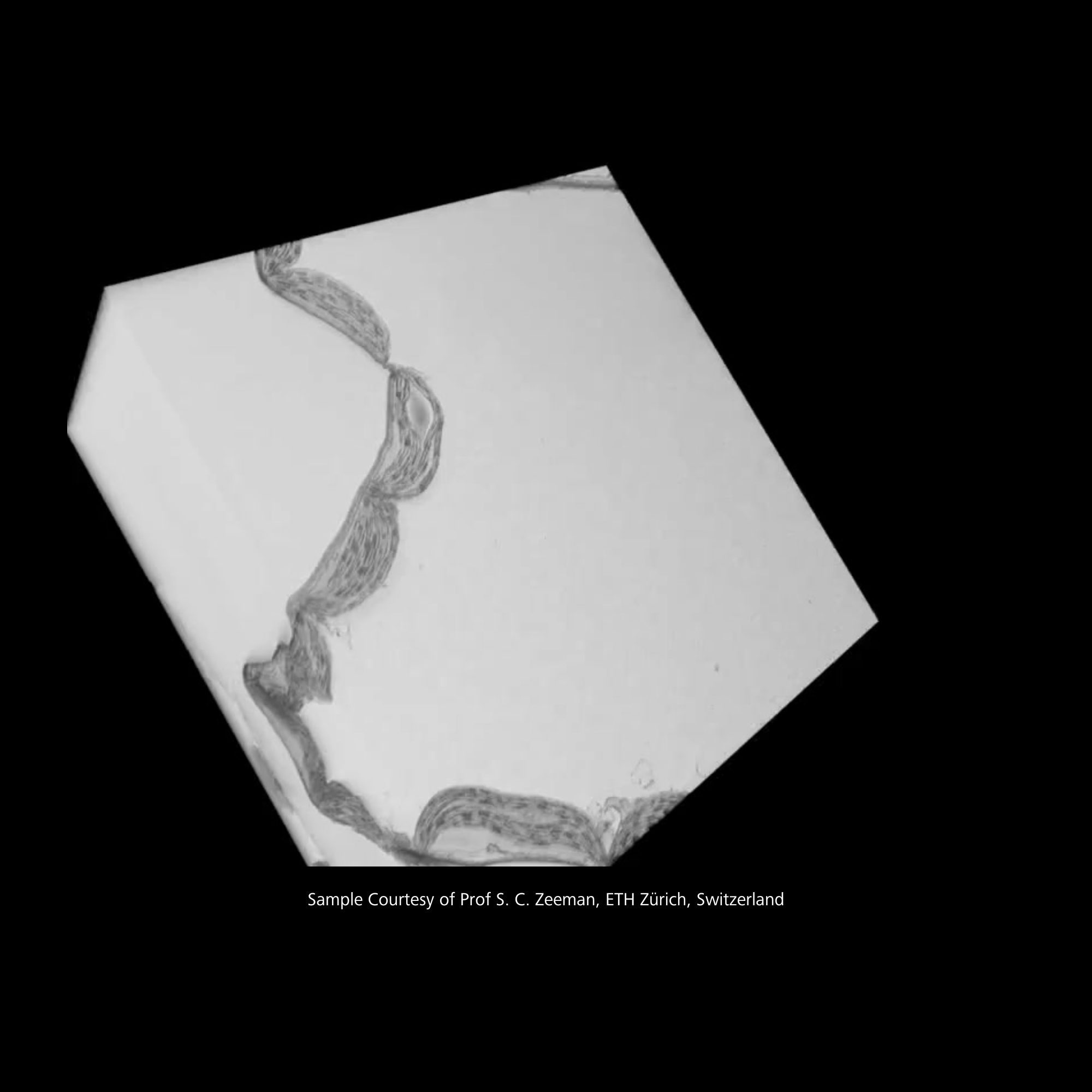
With the X-ray microscope, we can look at the entire structure at multiple scales and still with a very high resolution.
Recommended Products for Plant Research
Light-Sheet Multiview Imaging of Living and Cleared Specimens
ZEISS Lightsheet 7
Use ZEISS Lightsheet 7 to image large optically cleared specimens in toto – with subcellular resolution. Dedicated optics, sample chambers and holders allow adaption to the refractive index of your chosen clearing method.
Image large specimens in your preferred clearing solution
Get best image quality for various applications
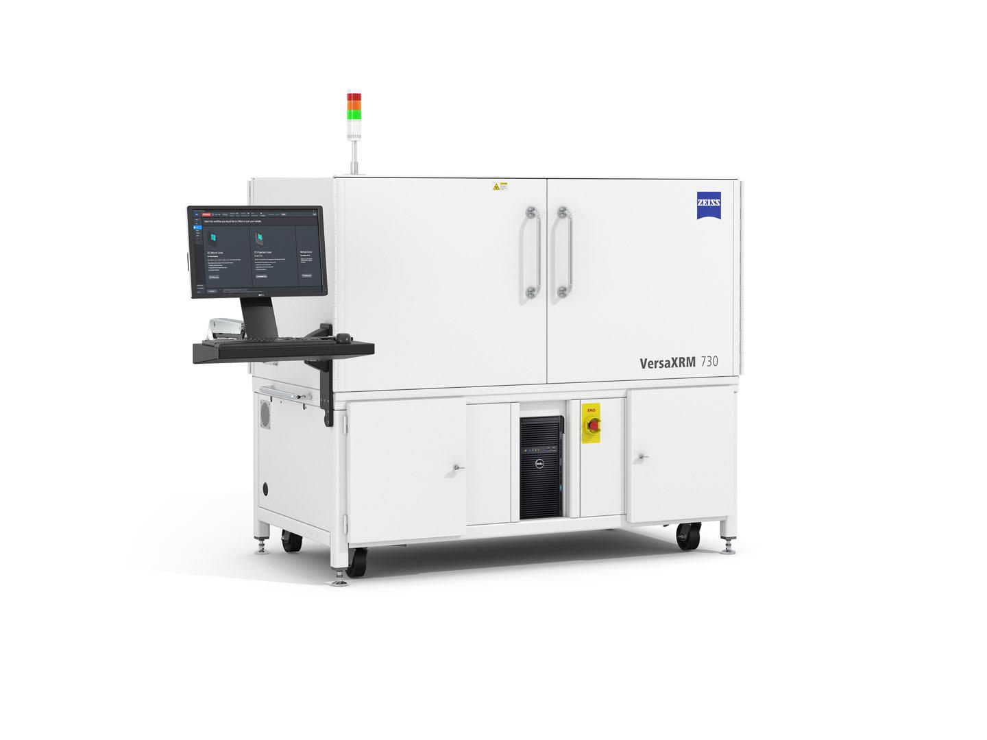
Discover More with 3D X-ray Imaging at Submicron Resolution
ZEISS Versa XRM
Experience the power of ZEISS Versa X-ray microscopes, the proven choice for researchers and scientists worldwide. Versa XRM feature intuitive user interfaces, ensuring that every user can maximize their productivity and achieve exceptional results.
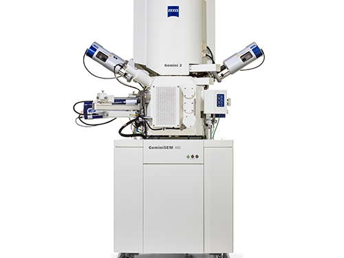
FE-SEM For Highest Demands in Sub-nanometer Imaging, Analytics and Sample Flexibility
ZEISS GeminiSEM
ZEISS GeminiSEM stands for effortless imaging with sub-nanometer resolution. These FE-SEMs (field emission scanning electron microscope) combine excellence in imaging and analytics. Innovations in electron optics and a new chamber design let you benefit from better image quality, usability and flexibility.
New standard for surface imaging – ZEISS GeminiSEM 560
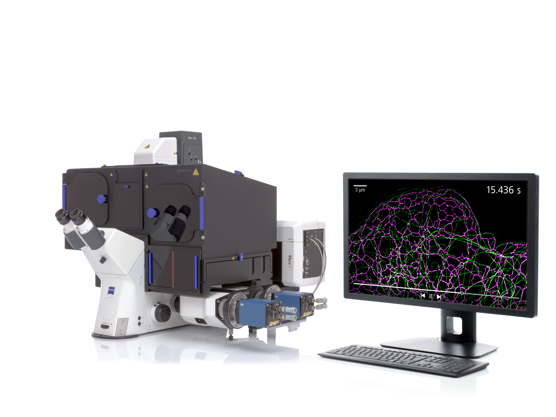
Live Imaging with Unprecedented Resolution down to Molecular Details
ZEISS Elyra 7
ZEISS Elyra 7 includes a wealth of microscopy techniques to meet your experimental needs across scales, optimally matching resolution, speed, and sensitivity requirements to your demanding samples.
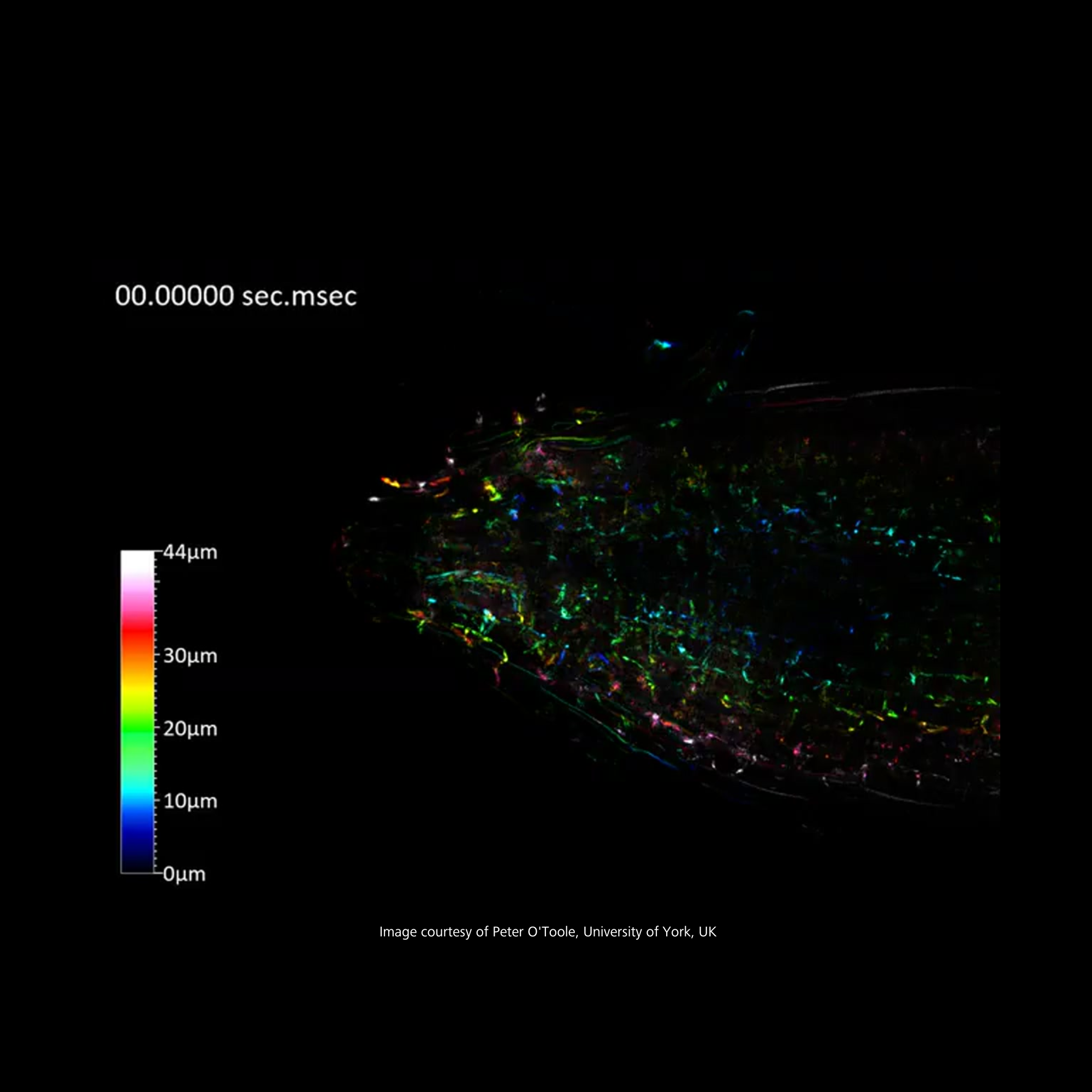
Instant Volumetric High-Speed Imaging of Living Organisms
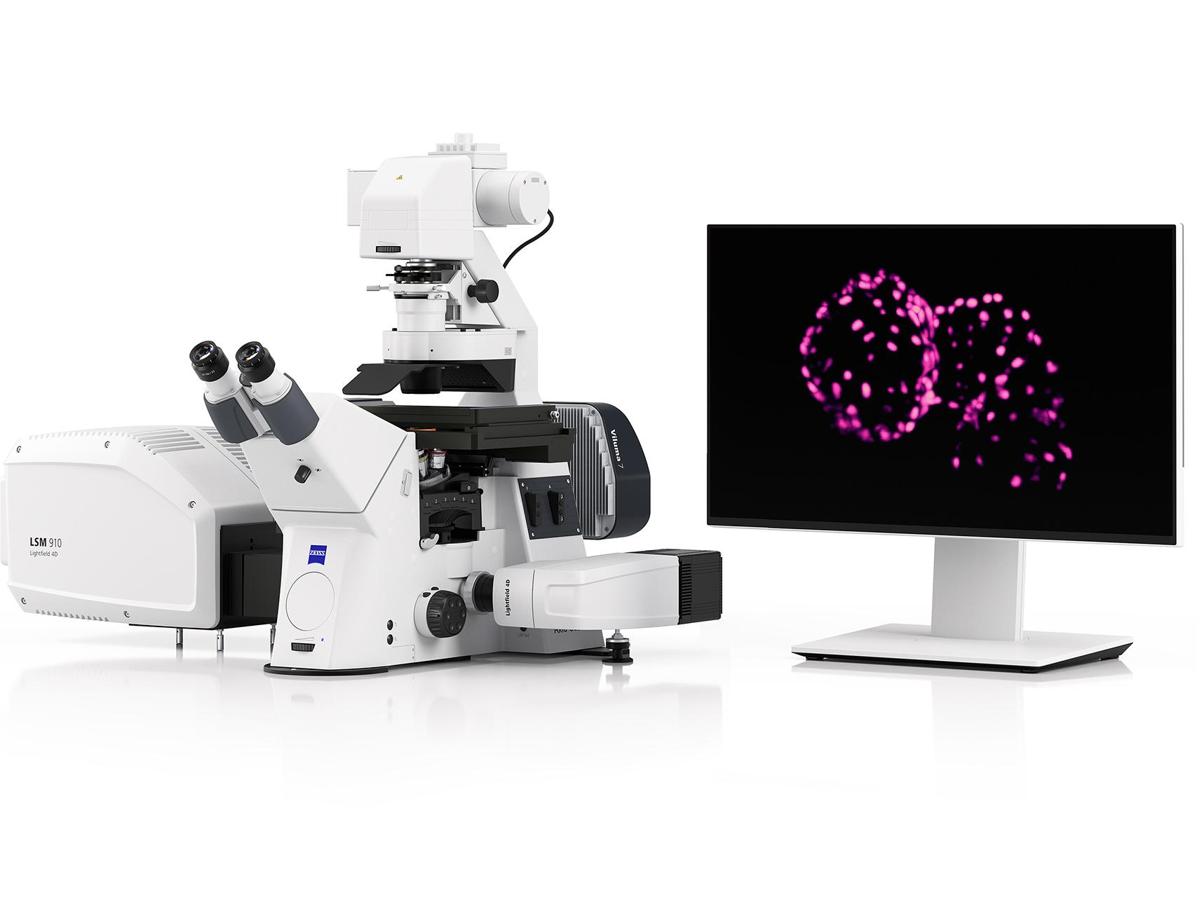
ZEISS LSM Lightfield 4D
Lightfield 4D is instant volumetric imaging at high speed. Acquire comprehensive 3D information with a single snap and say goodbye to any time delay within an imaged volume. For the first time, capture the fastest movements within whole organisms at up to 80 volumes per second – with all spatiotemporal information intact.
Your Solution for Advanced Image Analysis and Visualization
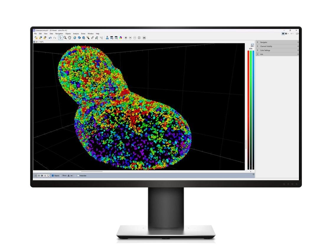
ZEISS arivis Pro
Maximize your image potential with our powerful image analysis and visualization tools. Create seamless analysis pipelines with just a few clicks. Effortlessly process massive datasets on compatible workstations.
ZEISS provides a comprehensive range of microscopes for plant biology, including:
Stereo and widefield microscopes for whole plant and tissue imaging
Laser scanning confocal and multiphoton microscopes for high-resolution, 3D imaging of plant cells and structure
Light-Sheet Microscopy for fast, gentle imaging of large specimens with minimal photobleaching, ideal for live plant imaging
Super-Resolution Microscopy for visualizing structures at a resolution beyond the diffraction limit of light, revealing fine details in plant cells
X-ray Tomography for non-destructive 3D imaging of internal plant structures, preserving the specimen's integrity
Volume Electron Microscopy (vEM) techniques for ultrastructural insights into plant tissues in 3D
ZEISS offers solutions, such as Light-Sheet Microscopy and Lattice Light-Sheet Microscopy, that allow researchers to observe dynamic processes in real-time. This capability is crucial for studying how plants respond to environmental stressors, such as drought or salinity, by visualizing cellular changes and adaptations as they occur.
Yes, ZEISS microscopes, particularly the LSM 980, utilize spectral imaging capabilities that enable the simultaneous imaging of multiple fluorescent markers while effectively separating autofluorescence. This is essential for studying complex cellular interactions in plant tissues without interference from naturally fluorescing compounds.
How does ZEISS support long-term time-lapse imaging of plant specimens?
ZEISS microscopy solutions, like Light-Sheet Microscopy, are designed for long-term time-lapse imaging by providing a stable environment with controlled conditions. This allows researchers to monitor dynamic processes, such as cell division and organ development, over extended periods while maintaining sample viability.
How can ZEISS software help with plant image analysis?
Data analysis is crucial for extracting meaningful insights from complex plant imaging datasets. ZEISS offers advanced software solutions, such as arivis Cloud and arivis Pro, which utilize machine learning for efficient cell segmentation and counting, phenotypic trait quantification and 3D reconstruction of plant structures. These automated, AI-driven tools streamline image analysis, enabling researchers to focus on their findings and discoveries.
How can ZEISS microscopy techniques enhance our understanding of photosynthesis and chloroplast function?
ZEISS offers a variety of microscopy solutions, such as super-resolution microscopy and fluorescence microscopy, to enable detailed visualization of chloroplast structure and function, pigment composition and stress responses and photosynthetic efficiency and energy transfer. By capturing high-resolution images and dynamic processes, researchers can uncover the mechanisms of photosynthesis and how plants optimize energy production under varying environmental conditions.