


Biopharma insights hub
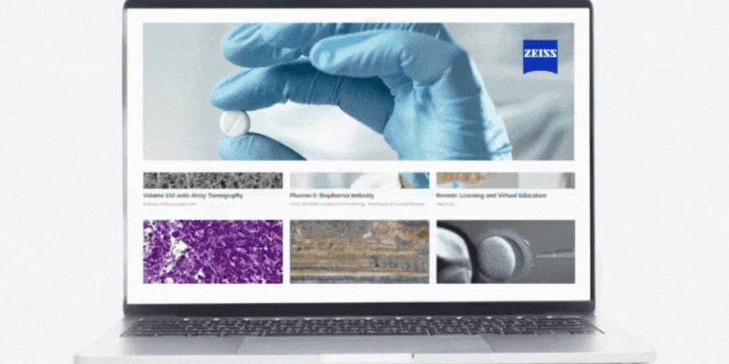
Discover tailored applications
Explore applications and discover tailored solutions for you industry needs.

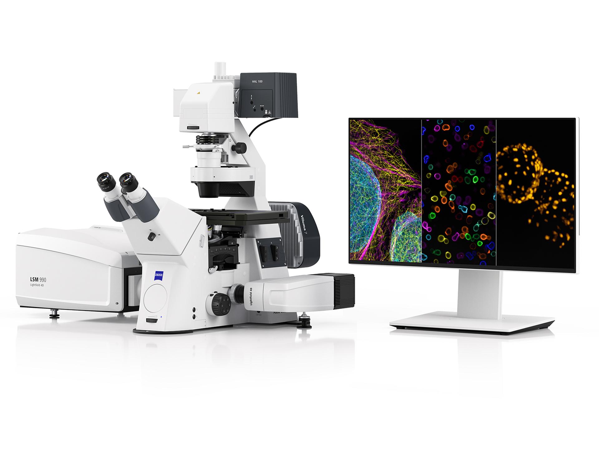
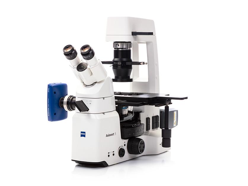
Our microscopy solutions push the boundaries of biotech, pharma, and industrial research by enabling high-resolution microscopy for druggable target identification, automated high-content screening for hit-to-lead selection and AI-driven imaging workflows.
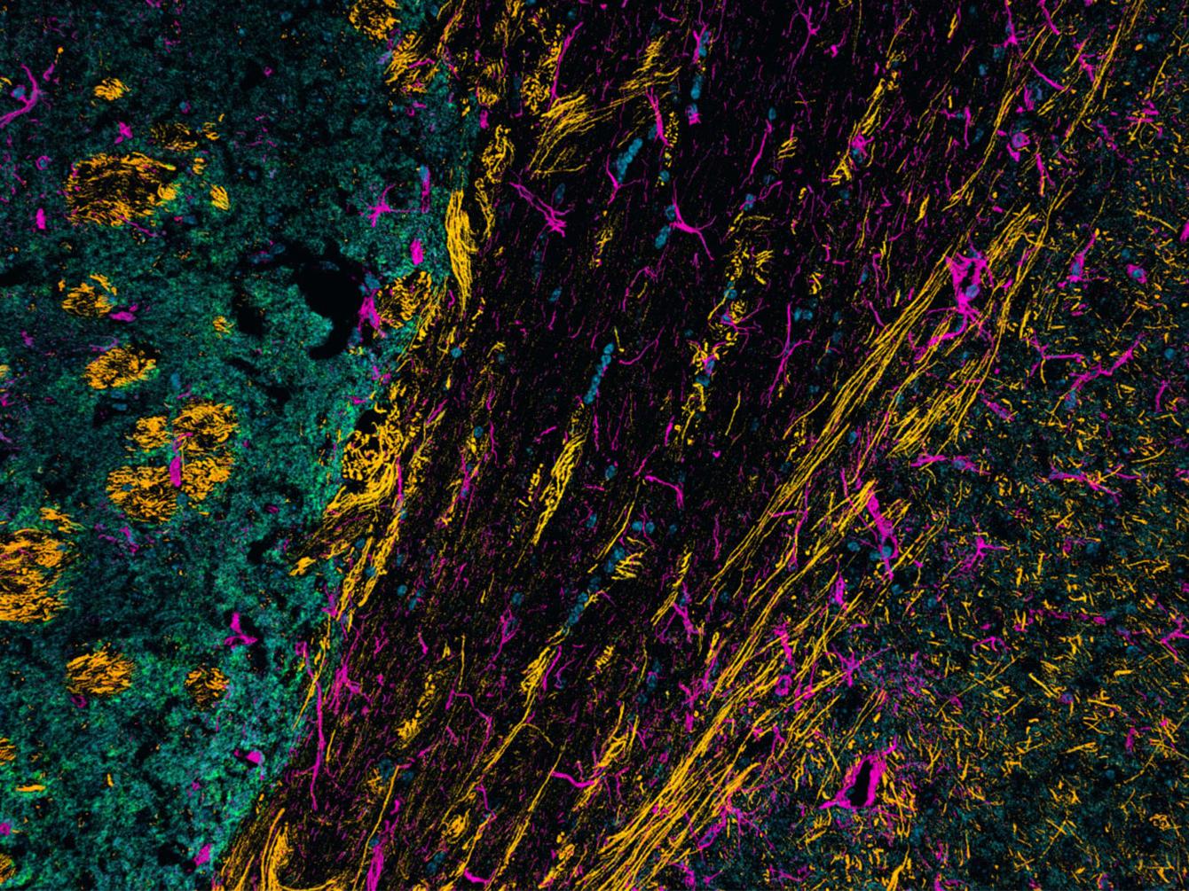
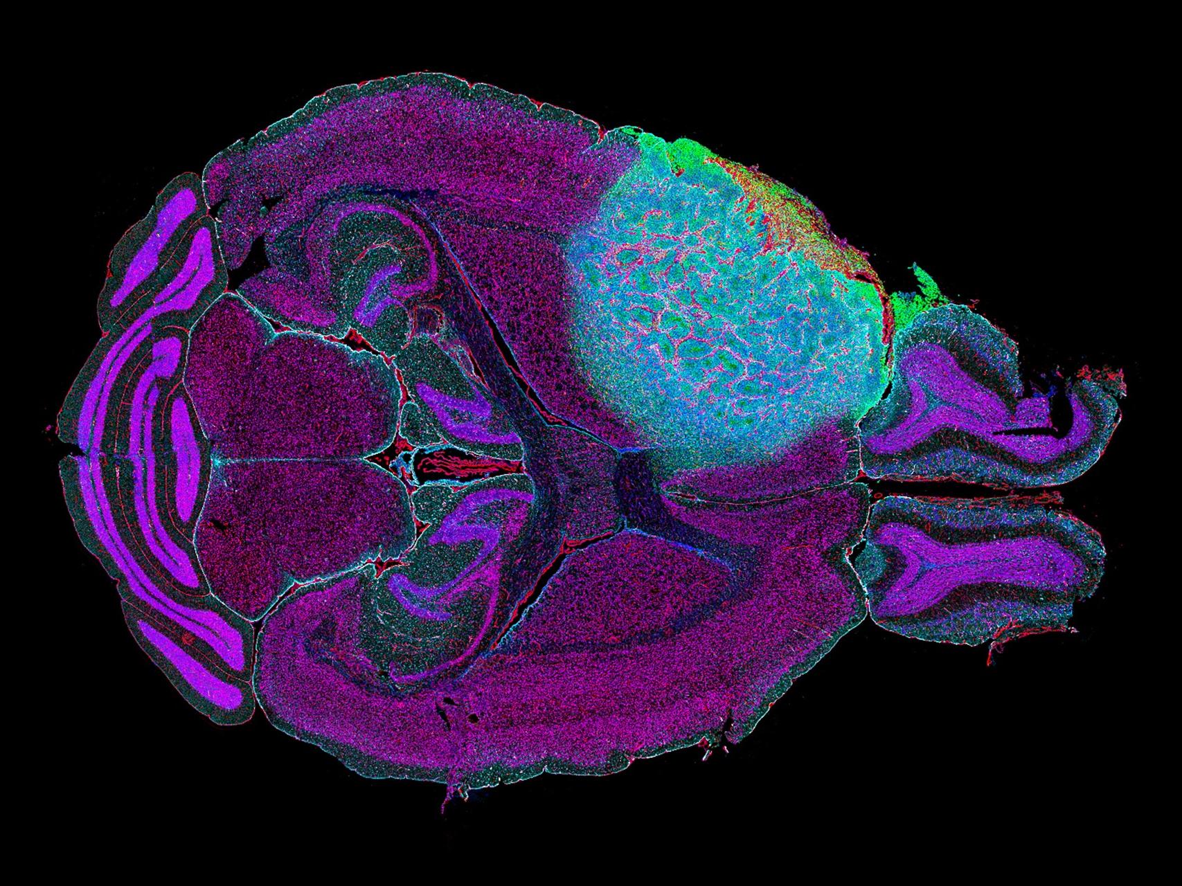
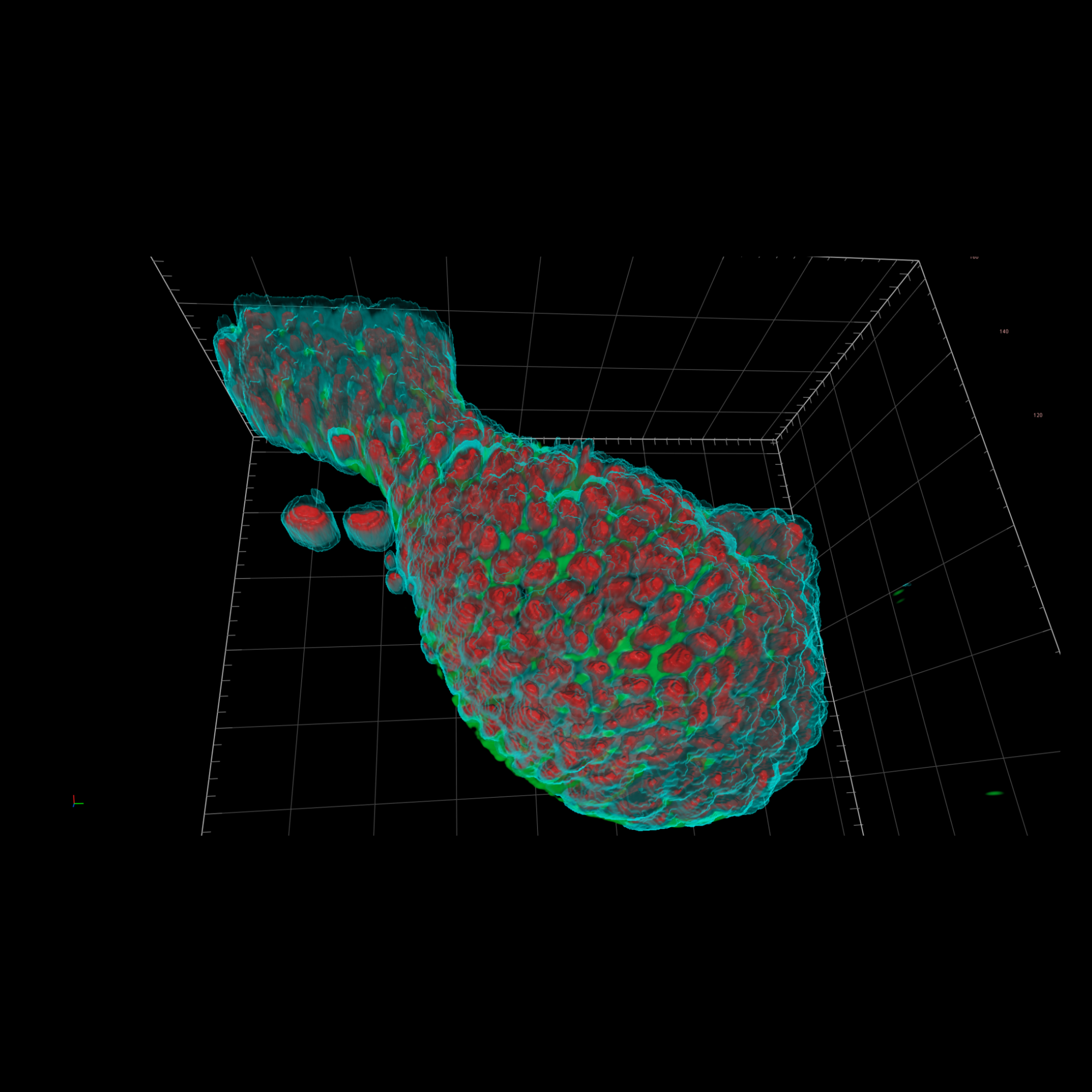
From 3D-cellular imaging to tissue analysis for pre-clinical studies, our devices deliver the precision, scalability, and automation needed to drive regulatory-compliant drug discovery workflows.


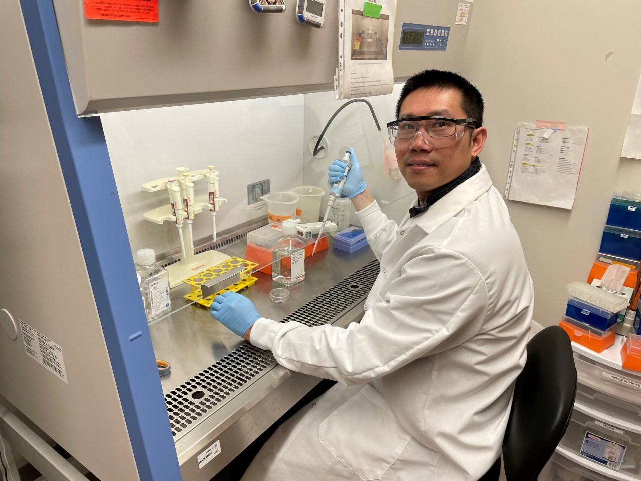


We are committed to empowering emerging biotech companies through our New Biotech Startup Program, partnering with leading incubators like Lab Central, BioLabs, JLabs, and CIC. We provide flexible imaging solutions, expert guidance, and scalable financing options to help startups drive innovation, generate high-quality data, and accelerate their path to discovery.


✔ Super-resolution imaging for identifying druggable targets & protein interactions
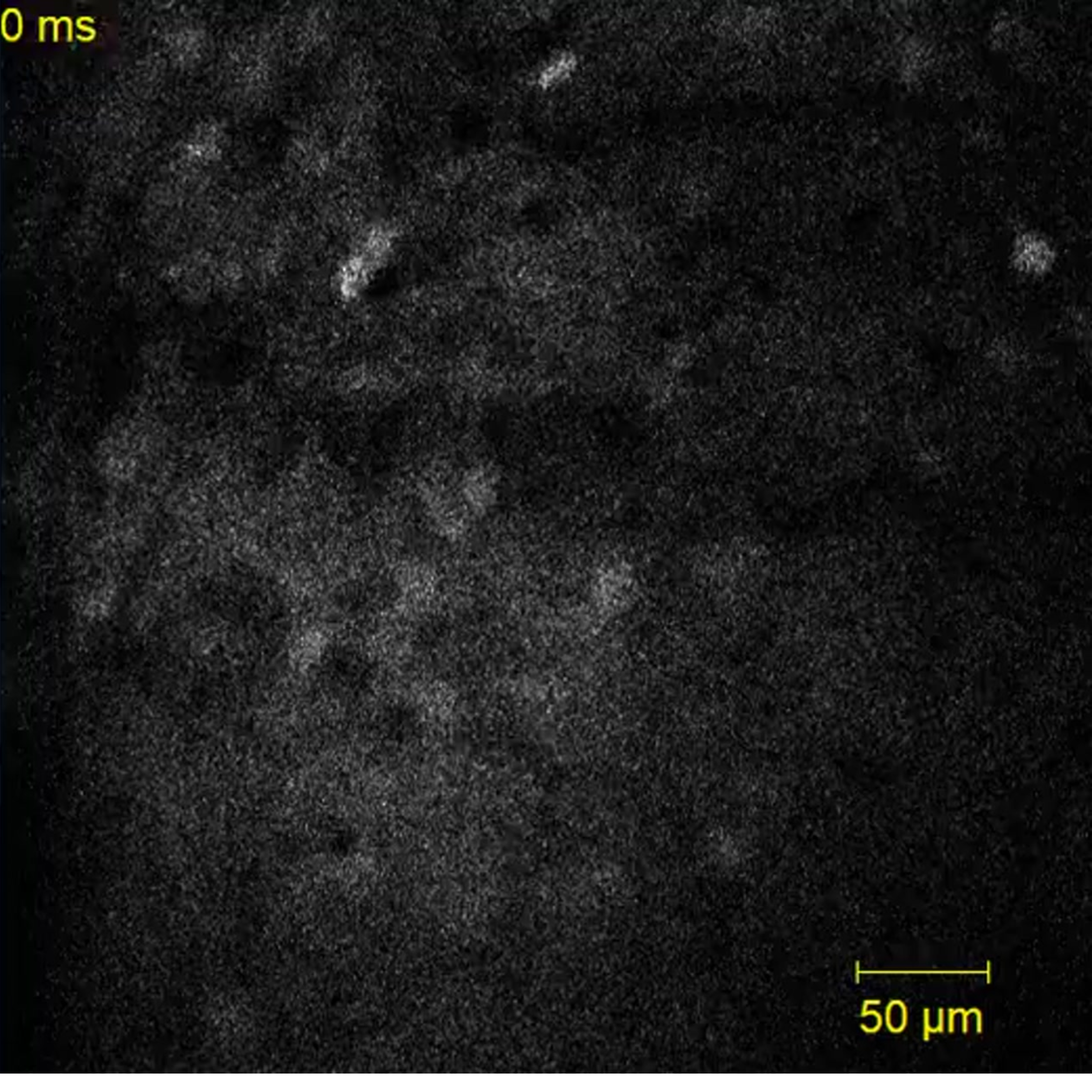
✔ Live-cell imaging for CRISPR screening & gene editing research
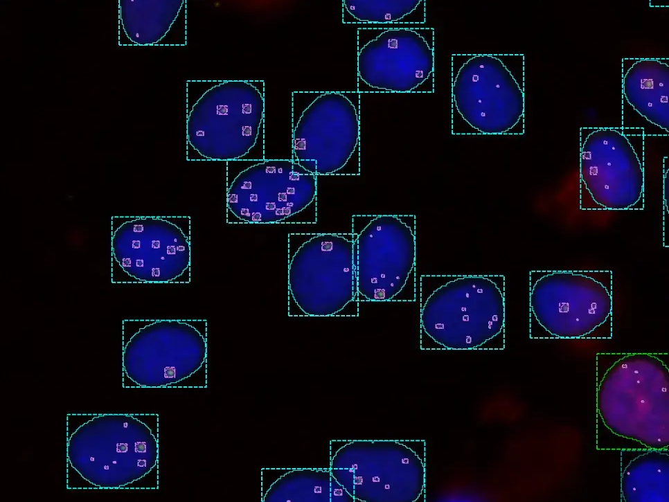
✔ AI-powered segmentation for predictive drug response modeling
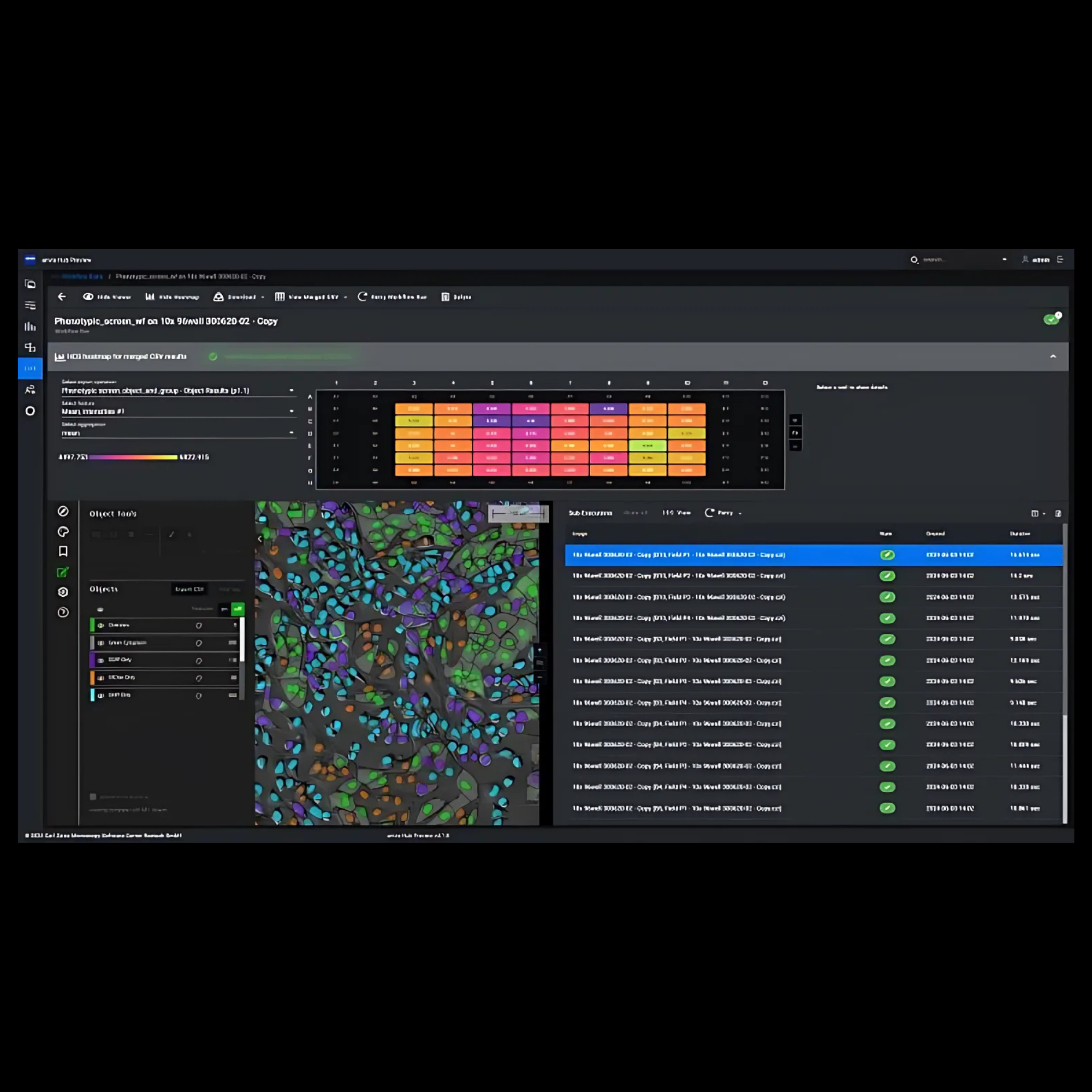

✔ High-content screening (HCS) with automated AI-driven microscopy
✔ Live-cell imaging for pharmacokinetics (PK) & dose-response modeling
✔ Automated imaging workflows for drug screening scalability
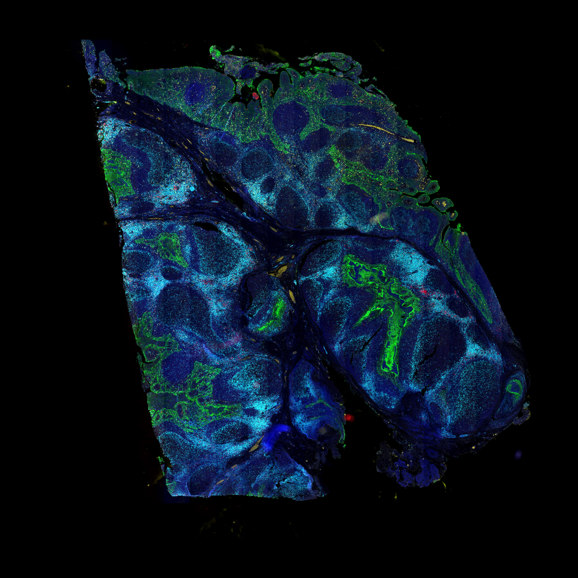

✔ AI-driven image analysis for spatial-omics & immuno-oncology
✔ Reproducible and easy to use imaging solutions for CROs
✔ Go from samples to finished reports


