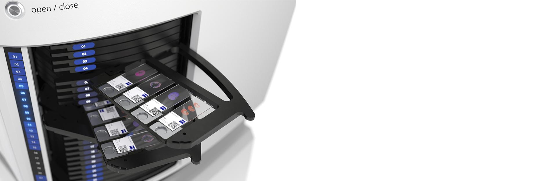

Digital Slide Scanner
ZEISS Axioscan 7
Discover high-performance digital slide scanning tailored to your application needs. Whether your focus is spatial biology at scale, life science research, clinical applications, or geology, the Axioscan 7 slide scanning microscope offers advanced automation capabilities and exceptional image quality.
Slide scanning solutions for your field of application
Discover ZEISS Axioscan 7 configurations designed to meet your unique requirements


Axioscan 7
Scanning performance combined with application freedom
- Application flexibility in a multi-user environment
- Rapid switching between fluorescence, brightfield, and polarization
- Research-grade data quality for demanding fluorescence applications
- Access to numerous additional processing and analysis functions via ZEN


Axioscan 7 spatial biology
Scalable multipex imaging for routine applications
- Fastest multiplex imaging of up to eight fluorescence stained biomarkers
- Includes solutions for robust, automated tissue detection and to support hyper-plex cyclic imaging assays
- Optimized configuration for superior inter-day and inter-device reproducibility
- Complementary software offerings for lab-automation, LIMS integration and AI-based image analysis


Axioscan 7 geology
Thin section slide scanning for digitization of petrography data
- Rapid digitization of geological thin sections with brightfield and polarization imaging
- Generation of complete petrographic data
- Intuitive navigation through rich petrographic thin section datasets
- AI-based mineral classification and analysis
-
Scanning performance combined with application freedom
Axioscan 7 combines qualities that you would not expect to get in a slide scanner: high speed digitization and outstanding image quality plus an unrivaled variety of imaging modes are all available in a fully automated and easy to operate system. The most challenging research tasks are supported by powerful hardware and perfectly featured software. Give your imaging facility users the ability to capture virtual slides quickly and with consistently high quality, whether their applications require brightfield, fluorescence or polarization imaging.
A Variety of Super-fast Brightfield Imaging Modes
The newly designed condenser with its motorized modulator disk allows automatic switching between different brightfield imaging modes to adapt to the different requirements of your applications, opening a new range of experiments and modality combinations.
- Dramatically improved scan speeds in all brightfield imaging modes
- Better sample detection and focusing
- New phase and relief contrast options
- Circular and linear polarization now fully supported


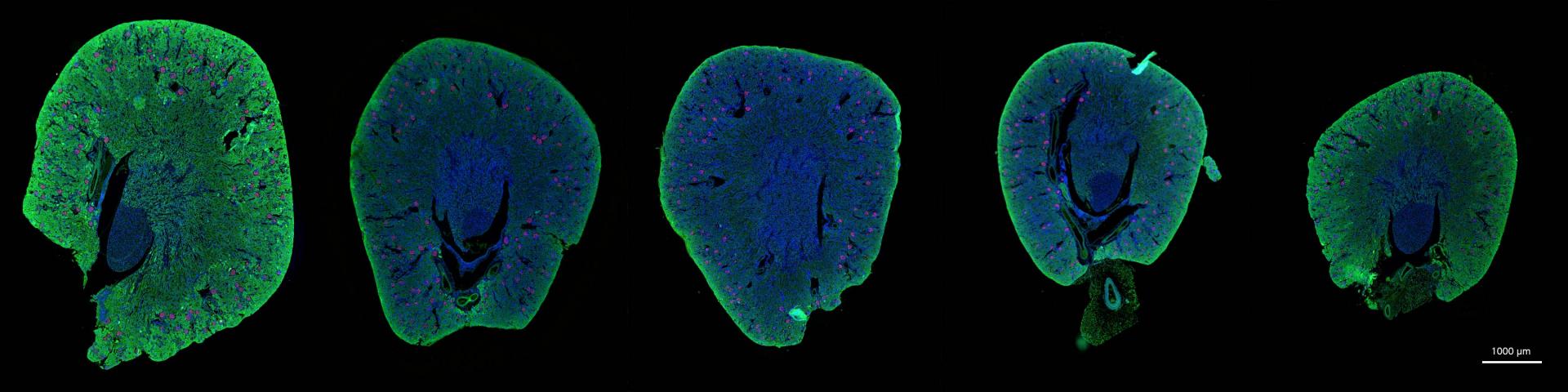
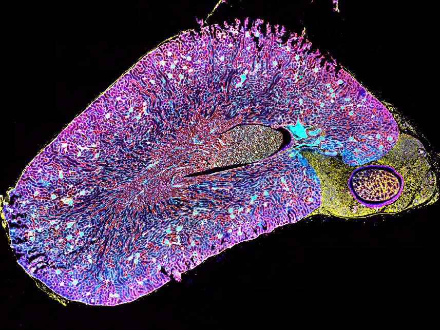
Paraffin-embedded mouse kidneys from healthy wildtype animals (12 weeks). Nephrin stained with Cy3. PCNA APC (FarRed) and DAPI as counterstaining. Imaged with 20× NA 0.8 objective.
Paraffin-embedded mouse kidneys from healthy wildtype animals (12 weeks). Nephrin stained with Cy3. PCNA APC (FarRed) and DAPI as counterstaining. Imaged with 20× NA 0.8 objective.

Solanum tuberosum – potatoe starch, 20x Plan-Apochromat 0.8; A) TIE phase contrast, B) TIE relief contrast, C) Brightfield
Solanum tuberosum – potatoe starch, 20x Plan-Apochromat 0.8; A) TIE phase contrast, B) TIE relief contrast, C) Brightfield
-
Workflow automation for multiplexed spatial profiling at scale
By leveraging multiplex immunofluorescence (mIF) staining with multiple biomarkers, spatial biology allows simultaneous visualization and quantification of numerous proteins within a single tissue section, enabling detailed analysis of cell presence, abundance, spatial distribution, and cell-to-cell interactions.
Load ZEISS Axioscan 7 spatial biology with 100 samples and scan them in less than a day with unprecedented speed, fully automated and supported by AI-enabled tissue detection and high dynamic range imaging. Analyze up to eight biomarkers at the same time and generate highly reproducible data you can rely on. We offer complimentary service solutions to integrate streamlined workflow solutions into pre-existing LIMS and IMS systems.


NSCLC tissue stained with UltiMapper I/O PD-L1 kit.


NSCLC tissue stained with UltiMapper I/O PD-L1 kit. Picture detail.
-
Thin section slide scanning for digitization of petrography data
Embrace digitalization with Axioscan 7, not only to create high-quality digitized petrography data efficiently, but also to gain the advantages of easy data sharing and seamless integration into your geological workflows. With AI-integrated analysis and remote collaboration capabilities, Axioscan 7 empowers geologists and researchers to work together seamlessly from anywhere in the world. Maximize the benefits of modern technology in quantitative petrography and automated analytics.


Circular Polarization
")
")
Cross Polarized Light (XPL)
")
")
Plane Polarized Light (PPL)


Mineral Phase Analysis
The combination of ZEISS Axioscan 7 and ZEISS AI-based segmentation enables automated analysis across large numbers of samples. Easy machine learning segmentation allows you to label each mineral phase of interest using an intuitive painting interface and allows the software to build a model of the mineralogy across your entire sample.


AI-Based Mineral Classification
Automated machine-learning based mineral classification using a single ZEN Intellesis model, applied here on two sandstone samples.
Both modal mineralogy and pore / grain sizes can be measured and automatically reported.

PPL-to-XPL
Full thin-section polarization images. This kyanite-bearing schist has been imaged as part of a digital thin section collection. The upper image shows a single PPL orientation, while the lower view shows the capture of the thin section in multiple XPL orientations. This allows a simulated stage rotation to observe and analyze extinction angles, with the full XPL variation over 90°.


PPL-to-Pleochroism
Close up of a single biotite grain within a granite sample. Sample has been imaged in multiple PPL orientations in order to observe the full range of pleochroism as the sample is rotated through 180° relative to the polarizer.

Correlative Microscopy
Use ZEN Connect to intuitively build correlative projects that start with the data-rich, light microscopy environment from ZEISS Axioscan 7 Geo. Here additional phase and geochemical information from ZEISS Mineralogic becomes the next step in a petrological investigation. Sample shown is a granulite facies metagabbro from Scouriemore, North West Scotland.


Mineral Orientation Analysis
The optional PetPAT orientation analysis package turns entire thin sections into powerful mineral orientation maps. These datasets can be used in conjunction with mineral segmentation for detailed studies and used to generate grain size distribution data.
- Use XPL image stacks to calculate the angle at which any given pixel is at maximum or minimum luminance
- Use these data to allow the entire thin section to be mapped for orientation of mineral grains in transmitted light


")
")
")
")


")
")
")
")


")
")
")
")