![arivis Pro arivis Pro]({"xsmall":"https://www.zeiss.com/content/dam/rms/reference-master/products/software/arivis-vision4d/arivis-pro-product.jpg/_jcr_content/renditions/original.image_file.100.100.156,46,848,738.file/arivis-pro-product.jpg","small":"https://www.zeiss.com/content/dam/rms/reference-master/products/software/arivis-vision4d/arivis-pro-product.jpg/_jcr_content/renditions/original.image_file.360.360.156,46,848,738.file/arivis-pro-product.jpg","medium":"https://www.zeiss.com/content/dam/rms/reference-master/products/software/arivis-vision4d/arivis-pro-product.jpg/_jcr_content/renditions/original.image_file.692.692.156,46,848,738.file/arivis-pro-product.jpg","large":"https://www.zeiss.com/content/dam/rms/reference-master/products/software/arivis-vision4d/arivis-pro-product.jpg/_jcr_content/renditions/original.image_file.692.692.156,46,848,738.file/arivis-pro-product.jpg","xlarge":"https://www.zeiss.com/content/dam/rms/reference-master/products/software/arivis-vision4d/arivis-pro-product.jpg/_jcr_content/renditions/original.image_file.692.692.156,46,848,738.file/arivis-pro-product.jpg","xxlarge":"https://www.zeiss.com/content/dam/rms/reference-master/products/software/arivis-vision4d/arivis-pro-product.jpg/_jcr_content/renditions/original.image_file.692.692.156,46,848,738.file/arivis-pro-product.jpg","max":"https://www.zeiss.com/content/dam/rms/reference-master/products/software/arivis-vision4d/arivis-pro-product.jpg/_jcr_content/renditions/original.image_file.692.692.156,46,848,738.file/arivis-pro-product.jpg"})
ZEISS arivis Pro
Your Solution for Advanced Image Analysis and Visualization
Maximize your image potential with our powerful image analysis and visualization tools. Create seamless analysis pipelines with just a few clicks. Effortlessly process massive datasets on compatible workstations. “Walk into your sample” using our VR toolkit for a new perspective.
![ZEISS arivis Pro Software for end-to-end, flexible pipelines ZEISS arivis Pro Software for end-to-end, flexible pipelines]({"xsmall":"https://www.zeiss.com/content/dam/rms/reference-master/products/software/arivis-vision4d/arivis-pro-strip.jpg/_jcr_content/renditions/original.image_file.100.100.file/arivis-pro-strip.jpg","small":"https://www.zeiss.com/content/dam/rms/reference-master/products/software/arivis-vision4d/arivis-pro-strip.jpg/_jcr_content/renditions/original.image_file.360.360.file/arivis-pro-strip.jpg","medium":"https://www.zeiss.com/content/dam/rms/reference-master/products/software/arivis-vision4d/arivis-pro-strip.jpg/_jcr_content/renditions/original.image_file.768.768.file/arivis-pro-strip.jpg","large":"https://www.zeiss.com/content/dam/rms/reference-master/products/software/arivis-vision4d/arivis-pro-strip.jpg/_jcr_content/renditions/original./arivis-pro-strip.jpg","xlarge":"https://www.zeiss.com/content/dam/rms/reference-master/products/software/arivis-vision4d/arivis-pro-strip.jpg/_jcr_content/renditions/original./arivis-pro-strip.jpg","xxlarge":"https://www.zeiss.com/content/dam/rms/reference-master/products/software/arivis-vision4d/arivis-pro-strip.jpg/_jcr_content/renditions/original./arivis-pro-strip.jpg","max":"https://www.zeiss.com/content/dam/rms/reference-master/products/software/arivis-vision4d/arivis-pro-strip.jpg/_jcr_content/renditions/original./arivis-pro-strip.jpg"})
Flexible, End-to-End Image Analysis Pipelines
ZEISS arivis Pro empowers you to automate image analysis and visualization pipelines. Leverage traditional methods or AI models effortlessly to create pipelines for any image size, dimension, or modality without the need to code.
The software supports and handles over 30 commercial file formats. Efficiently process large files with ease. Pre-configured pipelines and standard assays are available for both simple and demanding analysis tasks. Or you can customize pipelines for your specific goals.
It takes just one click to repeat your analysis for consistent, quantitative results. Boost productivity and ensure reproducible results
![ZEISS arivis Pro VR Toolkit for an immersive sample analysis ZEISS arivis Pro VR Toolkit for an immersive sample analysis]({"xsmall":"https://www.zeiss.com/content/dam/rms/reference-master/products/software/arivis-vision4d/octopus-gui-16x9.jpg/_jcr_content/renditions/original.image_file.100.56.file/octopus-gui-16x9.jpg","small":"https://www.zeiss.com/content/dam/rms/reference-master/products/software/arivis-vision4d/octopus-gui-16x9.jpg/_jcr_content/renditions/original.image_file.360.203.file/octopus-gui-16x9.jpg","medium":"https://www.zeiss.com/content/dam/rms/reference-master/products/software/arivis-vision4d/octopus-gui-16x9.jpg/_jcr_content/renditions/original.image_file.768.432.file/octopus-gui-16x9.jpg","large":"https://www.zeiss.com/content/dam/rms/reference-master/products/software/arivis-vision4d/octopus-gui-16x9.jpg/_jcr_content/renditions/original.image_file.1024.576.file/octopus-gui-16x9.jpg","xlarge":"https://www.zeiss.com/content/dam/rms/reference-master/products/software/arivis-vision4d/octopus-gui-16x9.jpg/_jcr_content/renditions/original.image_file.1280.720.file/octopus-gui-16x9.jpg","xxlarge":"https://www.zeiss.com/content/dam/rms/reference-master/products/software/arivis-vision4d/octopus-gui-16x9.jpg/_jcr_content/renditions/original.image_file.1440.810.file/octopus-gui-16x9.jpg","max":"https://www.zeiss.com/content/dam/rms/reference-master/products/software/arivis-vision4d/octopus-gui-16x9.jpg/_jcr_content/renditions/original./octopus-gui-16x9.jpg"})
ZEISS arivis Pro VR Toolkit for an immersive sample analysis
![]({"xsmall":"https://www.zeiss.com/content/dam/rms/reference-master/products/software/arivis-vision4d/octopus-gui-16x9.jpg/_jcr_content/renditions/original.image_file.100.56.file/octopus-gui-16x9.jpg","small":"https://www.zeiss.com/content/dam/rms/reference-master/products/software/arivis-vision4d/octopus-gui-16x9.jpg/_jcr_content/renditions/original.image_file.360.203.file/octopus-gui-16x9.jpg","medium":"https://www.zeiss.com/content/dam/rms/reference-master/products/software/arivis-vision4d/octopus-gui-16x9.jpg/_jcr_content/renditions/original.image_file.768.432.file/octopus-gui-16x9.jpg","large":"https://www.zeiss.com/content/dam/rms/reference-master/products/software/arivis-vision4d/octopus-gui-16x9.jpg/_jcr_content/renditions/original.image_file.1024.576.file/octopus-gui-16x9.jpg","xlarge":"https://www.zeiss.com/content/dam/rms/reference-master/products/software/arivis-vision4d/octopus-gui-16x9.jpg/_jcr_content/renditions/original.image_file.1280.720.file/octopus-gui-16x9.jpg","xxlarge":"https://www.zeiss.com/content/dam/rms/reference-master/products/software/arivis-vision4d/octopus-gui-16x9.jpg/_jcr_content/renditions/original.image_file.1440.810.file/octopus-gui-16x9.jpg","max":"https://www.zeiss.com/content/dam/rms/reference-master/products/software/arivis-vision4d/octopus-gui-16x9.jpg/_jcr_content/renditions/original./octopus-gui-16x9.jpg"})
ZEISS arivis Pro VR Toolkit
Get a new perspective on your sample with an immersive virtual reality experience. Freed from being tethered to your keyboard and mouse, you can directly move, rotate, scale, and shape your digital image data. Experience an in-depth perception equivalent to the real world.
- Explore your sample in 3D
- Observe details from diverse angles
- Collaborate with colleagues for a live review of your sample and results
- Easy voice and hands-on control
- Effective, interactive proofreading and editing of automatic analysis results.
![Sub cellular analysis based in AI model of FIB SEM image Sub cellular analysis based in AI model of FIB SEM image]({"xsmall":"https://www.zeiss.com/content/dam/rms/reference-master/products/software/arivis-vision4d/hela-crossbeam-highres.jpg/_jcr_content/renditions/original.image_file.100.56.file/hela-crossbeam-highres.jpg","small":"https://www.zeiss.com/content/dam/rms/reference-master/products/software/arivis-vision4d/hela-crossbeam-highres.jpg/_jcr_content/renditions/original.image_file.360.203.file/hela-crossbeam-highres.jpg","medium":"https://www.zeiss.com/content/dam/rms/reference-master/products/software/arivis-vision4d/hela-crossbeam-highres.jpg/_jcr_content/renditions/original.image_file.768.432.file/hela-crossbeam-highres.jpg","large":"https://www.zeiss.com/content/dam/rms/reference-master/products/software/arivis-vision4d/hela-crossbeam-highres.jpg/_jcr_content/renditions/original.image_file.1024.576.file/hela-crossbeam-highres.jpg","xlarge":"https://www.zeiss.com/content/dam/rms/reference-master/products/software/arivis-vision4d/hela-crossbeam-highres.jpg/_jcr_content/renditions/original.image_file.1280.720.file/hela-crossbeam-highres.jpg","xxlarge":"https://www.zeiss.com/content/dam/rms/reference-master/products/software/arivis-vision4d/hela-crossbeam-highres.jpg/_jcr_content/renditions/original.image_file.1440.810.file/hela-crossbeam-highres.jpg","max":"https://www.zeiss.com/content/dam/rms/reference-master/products/software/arivis-vision4d/hela-crossbeam-highres.jpg/_jcr_content/renditions/original./hela-crossbeam-highres.jpg"})
Sub cellular analysis based in AI model of FIB SEM image
![Sub cellular analysis based in AI model of FIB SEM image Sub cellular analysis based in AI model of FIB SEM image]({"xsmall":"https://www.zeiss.com/content/dam/rms/reference-master/products/software/arivis-vision4d/hela-crossbeam-highres.jpg/_jcr_content/renditions/original.image_file.100.56.file/hela-crossbeam-highres.jpg","small":"https://www.zeiss.com/content/dam/rms/reference-master/products/software/arivis-vision4d/hela-crossbeam-highres.jpg/_jcr_content/renditions/original.image_file.360.203.file/hela-crossbeam-highres.jpg","medium":"https://www.zeiss.com/content/dam/rms/reference-master/products/software/arivis-vision4d/hela-crossbeam-highres.jpg/_jcr_content/renditions/original.image_file.768.432.file/hela-crossbeam-highres.jpg","large":"https://www.zeiss.com/content/dam/rms/reference-master/products/software/arivis-vision4d/hela-crossbeam-highres.jpg/_jcr_content/renditions/original.image_file.1024.576.file/hela-crossbeam-highres.jpg","xlarge":"https://www.zeiss.com/content/dam/rms/reference-master/products/software/arivis-vision4d/hela-crossbeam-highres.jpg/_jcr_content/renditions/original.image_file.1280.720.file/hela-crossbeam-highres.jpg","xxlarge":"https://www.zeiss.com/content/dam/rms/reference-master/products/software/arivis-vision4d/hela-crossbeam-highres.jpg/_jcr_content/renditions/original.image_file.1440.810.file/hela-crossbeam-highres.jpg","max":"https://www.zeiss.com/content/dam/rms/reference-master/products/software/arivis-vision4d/hela-crossbeam-highres.jpg/_jcr_content/renditions/original./hela-crossbeam-highres.jpg"})
Advanced 3D Analysis
Boost results quickly with AI, even for complex tasks. Annotate slices in your Z-stack to train your model segmentation and classification and then apply it to the whole dataset. Use Machine Learning and Deep Learning operators locally or train models in the cloud and then import them to form the core of your automated pipeline. Visualize results in 3D and create videos for a polished analysis report with a few clicks.
![3D organoid analysis 3D organoid analysis]({"xsmall":"https://www.zeiss.com/content/dam/rms/reference-master/products/software/arivis-vision4d/organoid3.jpg/_jcr_content/renditions/original.image_file.100.56.file/organoid3.jpg","small":"https://www.zeiss.com/content/dam/rms/reference-master/products/software/arivis-vision4d/organoid3.jpg/_jcr_content/renditions/original.image_file.360.203.file/organoid3.jpg","medium":"https://www.zeiss.com/content/dam/rms/reference-master/products/software/arivis-vision4d/organoid3.jpg/_jcr_content/renditions/original.image_file.768.432.file/organoid3.jpg","large":"https://www.zeiss.com/content/dam/rms/reference-master/products/software/arivis-vision4d/organoid3.jpg/_jcr_content/renditions/original.image_file.1024.576.file/organoid3.jpg","xlarge":"https://www.zeiss.com/content/dam/rms/reference-master/products/software/arivis-vision4d/organoid3.jpg/_jcr_content/renditions/original.image_file.1280.720.file/organoid3.jpg","xxlarge":"https://www.zeiss.com/content/dam/rms/reference-master/products/software/arivis-vision4d/organoid3.jpg/_jcr_content/renditions/original.image_file.1440.810.file/organoid3.jpg","max":"https://www.zeiss.com/content/dam/rms/reference-master/products/software/arivis-vision4d/organoid3.jpg/_jcr_content/renditions/original./organoid3.jpg"})
3D organoid analysis
![]({"xsmall":"https://www.zeiss.com/content/dam/rms/reference-master/products/software/arivis-vision4d/organoid3.jpg/_jcr_content/renditions/original.image_file.100.56.file/organoid3.jpg","small":"https://www.zeiss.com/content/dam/rms/reference-master/products/software/arivis-vision4d/organoid3.jpg/_jcr_content/renditions/original.image_file.360.203.file/organoid3.jpg","medium":"https://www.zeiss.com/content/dam/rms/reference-master/products/software/arivis-vision4d/organoid3.jpg/_jcr_content/renditions/original.image_file.768.432.file/organoid3.jpg","large":"https://www.zeiss.com/content/dam/rms/reference-master/products/software/arivis-vision4d/organoid3.jpg/_jcr_content/renditions/original.image_file.1024.576.file/organoid3.jpg","xlarge":"https://www.zeiss.com/content/dam/rms/reference-master/products/software/arivis-vision4d/organoid3.jpg/_jcr_content/renditions/original.image_file.1280.720.file/organoid3.jpg","xxlarge":"https://www.zeiss.com/content/dam/rms/reference-master/products/software/arivis-vision4d/organoid3.jpg/_jcr_content/renditions/original.image_file.1440.810.file/organoid3.jpg","max":"https://www.zeiss.com/content/dam/rms/reference-master/products/software/arivis-vision4d/organoid3.jpg/_jcr_content/renditions/original./organoid3.jpg"})
High Content Analysis
Complex disease drug discovery demands advanced image analysis methods such as screening against a broader view of cell or tissue phenotype, scalable from 2D to 4D. Train an AI model using ZEISS arivis software on the cloud or locally, and automate image analysis to save time, reduce human bias, and gain consistent results.
![Tracking with ZEISS arivis Pro VR Tracking with ZEISS arivis Pro VR]({"xsmall":"https://www.zeiss.com/content/dam/rms/reference-master/products/software/arivis-vision4d/vr-track.jpg/_jcr_content/renditions/original.image_file.100.56.0,0,1470,827.file/vr-track.jpg","small":"https://www.zeiss.com/content/dam/rms/reference-master/products/software/arivis-vision4d/vr-track.jpg/_jcr_content/renditions/original.image_file.360.203.0,0,1470,827.file/vr-track.jpg","medium":"https://www.zeiss.com/content/dam/rms/reference-master/products/software/arivis-vision4d/vr-track.jpg/_jcr_content/renditions/original.image_file.768.432.0,0,1470,827.file/vr-track.jpg","large":"https://www.zeiss.com/content/dam/rms/reference-master/products/software/arivis-vision4d/vr-track.jpg/_jcr_content/renditions/original.image_file.1024.576.0,0,1470,827.file/vr-track.jpg","xlarge":"https://www.zeiss.com/content/dam/rms/reference-master/products/software/arivis-vision4d/vr-track.jpg/_jcr_content/renditions/original.image_file.1280.720.0,0,1470,827.file/vr-track.jpg","xxlarge":"https://www.zeiss.com/content/dam/rms/reference-master/products/software/arivis-vision4d/vr-track.jpg/_jcr_content/renditions/original.image_file.1440.810.0,0,1470,827.file/vr-track.jpg","max":"https://www.zeiss.com/content/dam/rms/reference-master/products/software/arivis-vision4d/vr-track.jpg/_jcr_content/renditions/original.image_file.1470.827.0,0,1470,827.file/vr-track.jpg"})
Tracking with ZEISS arivis Pro VR
![Tracking with ZEISS arivis Pro VR Tracking with ZEISS arivis Pro VR]({"xsmall":"https://www.zeiss.com/content/dam/rms/reference-master/products/software/arivis-vision4d/vr-track.jpg/_jcr_content/renditions/original.image_file.100.56.file/vr-track.jpg","small":"https://www.zeiss.com/content/dam/rms/reference-master/products/software/arivis-vision4d/vr-track.jpg/_jcr_content/renditions/original.image_file.360.203.file/vr-track.jpg","medium":"https://www.zeiss.com/content/dam/rms/reference-master/products/software/arivis-vision4d/vr-track.jpg/_jcr_content/renditions/original.image_file.768.432.file/vr-track.jpg","large":"https://www.zeiss.com/content/dam/rms/reference-master/products/software/arivis-vision4d/vr-track.jpg/_jcr_content/renditions/original.image_file.1024.576.file/vr-track.jpg","xlarge":"https://www.zeiss.com/content/dam/rms/reference-master/products/software/arivis-vision4d/vr-track.jpg/_jcr_content/renditions/original.image_file.1280.720.file/vr-track.jpg","xxlarge":"https://www.zeiss.com/content/dam/rms/reference-master/products/software/arivis-vision4d/vr-track.jpg/_jcr_content/renditions/original.image_file.1440.810.file/vr-track.jpg","max":"https://www.zeiss.com/content/dam/rms/reference-master/products/software/arivis-vision4d/vr-track.jpg/_jcr_content/renditions/original./vr-track.jpg"})
Tracking and Lineage
Quantify cell division and migratory phenotypes, and track changes in 2D and 3D image sets of any size. Use ZEISS arivis Pro VR to accurately proofread automatically generated tracks in an immersive VR environment. Easily reapply your pipeline to more datasets.
![Automated 3D neuron tracing of neuronal tissue (mouse brain). Automated 3D neuron tracing of neuronal tissue (mouse brain).]({"xsmall":"https://www.zeiss.com/content/dam/rms/reference-master/products/software/arivis-vision4d/forest-of-neurons-lsm980-arivis-vision4d.jpg/_jcr_content/renditions/original.image_file.100.56.file/forest-of-neurons-lsm980-arivis-vision4d.jpg","small":"https://www.zeiss.com/content/dam/rms/reference-master/products/software/arivis-vision4d/forest-of-neurons-lsm980-arivis-vision4d.jpg/_jcr_content/renditions/original.image_file.360.203.file/forest-of-neurons-lsm980-arivis-vision4d.jpg","medium":"https://www.zeiss.com/content/dam/rms/reference-master/products/software/arivis-vision4d/forest-of-neurons-lsm980-arivis-vision4d.jpg/_jcr_content/renditions/original.image_file.768.432.file/forest-of-neurons-lsm980-arivis-vision4d.jpg","large":"https://www.zeiss.com/content/dam/rms/reference-master/products/software/arivis-vision4d/forest-of-neurons-lsm980-arivis-vision4d.jpg/_jcr_content/renditions/original.image_file.1024.576.file/forest-of-neurons-lsm980-arivis-vision4d.jpg","xlarge":"https://www.zeiss.com/content/dam/rms/reference-master/products/software/arivis-vision4d/forest-of-neurons-lsm980-arivis-vision4d.jpg/_jcr_content/renditions/original.image_file.1280.720.file/forest-of-neurons-lsm980-arivis-vision4d.jpg","xxlarge":"https://www.zeiss.com/content/dam/rms/reference-master/products/software/arivis-vision4d/forest-of-neurons-lsm980-arivis-vision4d.jpg/_jcr_content/renditions/original.image_file.1440.810.file/forest-of-neurons-lsm980-arivis-vision4d.jpg","max":"https://www.zeiss.com/content/dam/rms/reference-master/products/software/arivis-vision4d/forest-of-neurons-lsm980-arivis-vision4d.jpg/_jcr_content/renditions/original./forest-of-neurons-lsm980-arivis-vision4d.jpg"})
Automated 3D neuron tracing of neuronal tissue (mouse brain).
![Automated 3D neuron tracing of neuronal tissue (mouse brain). Courtesy of Dr. Steffen Burgold, ZEISS RMS Customer Center Oberkochen, Germany. Automated 3D neuron tracing of neuronal tissue (mouse brain). Courtesy of Dr. Steffen Burgold, ZEISS RMS Customer Center Oberkochen, Germany.]({"xsmall":"https://www.zeiss.com/content/dam/rms/reference-master/products/software/arivis-vision4d/forest-of-neurons-lsm980-arivis-vision4d.jpg/_jcr_content/renditions/original.image_file.100.56.file/forest-of-neurons-lsm980-arivis-vision4d.jpg","small":"https://www.zeiss.com/content/dam/rms/reference-master/products/software/arivis-vision4d/forest-of-neurons-lsm980-arivis-vision4d.jpg/_jcr_content/renditions/original.image_file.360.203.file/forest-of-neurons-lsm980-arivis-vision4d.jpg","medium":"https://www.zeiss.com/content/dam/rms/reference-master/products/software/arivis-vision4d/forest-of-neurons-lsm980-arivis-vision4d.jpg/_jcr_content/renditions/original.image_file.768.432.file/forest-of-neurons-lsm980-arivis-vision4d.jpg","large":"https://www.zeiss.com/content/dam/rms/reference-master/products/software/arivis-vision4d/forest-of-neurons-lsm980-arivis-vision4d.jpg/_jcr_content/renditions/original.image_file.1024.576.file/forest-of-neurons-lsm980-arivis-vision4d.jpg","xlarge":"https://www.zeiss.com/content/dam/rms/reference-master/products/software/arivis-vision4d/forest-of-neurons-lsm980-arivis-vision4d.jpg/_jcr_content/renditions/original.image_file.1280.720.file/forest-of-neurons-lsm980-arivis-vision4d.jpg","xxlarge":"https://www.zeiss.com/content/dam/rms/reference-master/products/software/arivis-vision4d/forest-of-neurons-lsm980-arivis-vision4d.jpg/_jcr_content/renditions/original.image_file.1440.810.file/forest-of-neurons-lsm980-arivis-vision4d.jpg","max":"https://www.zeiss.com/content/dam/rms/reference-master/products/software/arivis-vision4d/forest-of-neurons-lsm980-arivis-vision4d.jpg/_jcr_content/renditions/original./forest-of-neurons-lsm980-arivis-vision4d.jpg"})
Neurobiology
Deploy cutting-edge neuron tracing algorithms for automatic, repeatable analysis of diverse images with a single click. Quantify neurons, glia/ microglia cells, axons, and more, even with low signal-to-noise ratio (SNR). Edit traces easily, using our semi-automatic interactive and VR tools. Analyze past experiment data, no matter the image format.
![Convert Over 30 Image File Formats Convert Over 30 Image File Formats]({"xsmall":"https://www.zeiss.com/content/dam/rms/reference-master/products/software/arivis-vision4d/adobestock-30-image-formats-16x9.jpg/_jcr_content/renditions/original.image_file.100.56.file/adobestock-30-image-formats-16x9.jpg","small":"https://www.zeiss.com/content/dam/rms/reference-master/products/software/arivis-vision4d/adobestock-30-image-formats-16x9.jpg/_jcr_content/renditions/original.image_file.360.203.file/adobestock-30-image-formats-16x9.jpg","medium":"https://www.zeiss.com/content/dam/rms/reference-master/products/software/arivis-vision4d/adobestock-30-image-formats-16x9.jpg/_jcr_content/renditions/original.image_file.768.432.file/adobestock-30-image-formats-16x9.jpg","large":"https://www.zeiss.com/content/dam/rms/reference-master/products/software/arivis-vision4d/adobestock-30-image-formats-16x9.jpg/_jcr_content/renditions/original.image_file.1024.576.file/adobestock-30-image-formats-16x9.jpg","xlarge":"https://www.zeiss.com/content/dam/rms/reference-master/products/software/arivis-vision4d/adobestock-30-image-formats-16x9.jpg/_jcr_content/renditions/original.image_file.1280.720.file/adobestock-30-image-formats-16x9.jpg","xxlarge":"https://www.zeiss.com/content/dam/rms/reference-master/products/software/arivis-vision4d/adobestock-30-image-formats-16x9.jpg/_jcr_content/renditions/original.image_file.1440.810.file/adobestock-30-image-formats-16x9.jpg","max":"https://www.zeiss.com/content/dam/rms/reference-master/products/software/arivis-vision4d/adobestock-30-image-formats-16x9.jpg/_jcr_content/renditions/original./adobestock-30-image-formats-16x9.jpg"})
Convert Over 30 Image File Formats
No matter what instrument you used to create your image, which format or file size: the ZEISS arivis SIS Converter easily converts your image file to our Scalable Image Storage (SIS) format, for fast and interactive exploration of your images with ZEISS arivis Pro, arivis Pro VR and arivis Hub. Save considerable time, while retaining all the data for advanced analysis.
- Supports file conversion for over 30 formats
- Que several import and conversion tasks
- Unattended batch conversion
- User-friendly file conversion task management interface
- Not only microscopy images (e.g., CT, MRI)
How Does It Work
Newest arivis Pro Release Highlights
Find out if you can benefit from the latest features available with the most current ZEISS arivis Pro release.
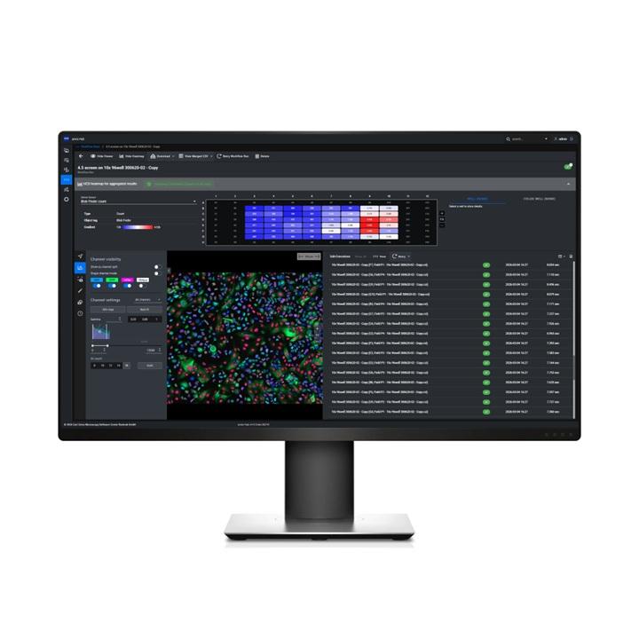
-
Summary of changes in arivis Vision4D 4.1.2 released 16 November 2023
Cellpose Segmenter:
- improved: better memory checks to avoid internal errors
- improved: more clear error and warnings
- improved: better description of the available pre-trained models in the help
- change: perform planewise is enabled by default now
- fix: handling of results for time points other than the first one (in combination with voxel filters)
- fix: normalization for data with bias in min intensity value
- fix: other small fixes for the Cellpose Segmenter
Import:
- improved: importing channel names for XDCE data
- fix: import problems for some ND2 time series
General:
- improved: faster free consumed license after quit of the application
-
Summary of changes in arivis Vision4D 4.1.1, released 25 July 2023
New & Noteworthy:
- Cellpose Segmenter - directly import pre-trained Cellpose models for easy and fast segmentation in the Analysis PipelineThis is a placeholder headline
Analysis: - new: native Cellpose Segmenter analysis operation (no Python needed)
- new: export a created model from Deep Learning Trainer as CZANN
- improved: Deep Learning - better compatibility with N2V models from ZEISS ZEN
- improved: Allow Holes option is on by default for the DL Segmenter
- improved: better conflict validation when renaming DL trainings
- fix: rare cases where positions of tracks after scaled analysis are incorrect
- fix: potential wrong segment reference for cell body in a trace
4D Viewer:
- improved: auto zoom when double clicking an object is more usable in 4D Viewer
- fix: sometimes traces (in cone display style) were not selectable in 4D Viewer
General:
- new: provide a description and an offline download for optional components in the installer
- fix: improved compatibility for DeltaVision DV import
- fix: What's New opens after an update more reliable
- fix: installer makes sure that no files from former Vision4D versions are left in the installation folder
- additional small fixes and improvements in the arivis_dl library
- Cellpose Segmenter - directly import pre-trained Cellpose models for easy and fast segmentation in the Analysis PipelineThis is a placeholder headline
-
Summary of changes in arivis Vision4D 4.1, released 27 March 2023
New & Noteworthy:
- New optional module: arivis AI toolkit features a complete Deep Learning workflow right in Vision4D - annotate, train & inference
- Various improvements in the tracing workflow
- Wellplate Editor to manually assign imagesets to wells or fields of a well
arivis AI Toolkit:
- new: Optional module arivis AI toolkit:
- new: Deep Learning Trainer panel to setup and train a DL model
- new: use the Draw Tool to draw segments to annotate regions for classes
- new: utilizing of sparse annotations to overcome the need to draw on every pixel
- new: drawn objects are sorted by class order - pixels are assigned to the topmost one (no need to cut out overlaps)
- new: add and reuse existing segments for class definition
- new: automatic clustering based on annotated backgound possible
- new: run and monitor the DL training process right from Vision4D
- new: directly open the trained DL model in the Analysis Pipeline for inference
- new: export the trained DL model as ONNX
- new: additional installer package to setup a whole DL Python environment
Tools:
- new: Draw Tool has a context menu and better keyboard shortcut support
- new: Draw Tool is aware of the DL Training panel
- new: Draw Tool allows to change class to draw by shortcut or context menu (in DL Trainer mode)
- improved: Trace Tool - connect to a cell body segment
- improved: Trace Tool - optimized remove branch
- improved: 'unable to connect' messages in the Trace Tool are more understandable
Analysis:
- new: 'Neuron Tracer' (from the Segmentation category) has been renamed to 'Neurite Tracer'
- new: 'Neuron Tracer (from Cell Body)' has been renamed to 'Neuron Tracer'
- new: Neuron Tracer preview shows all parts in the same color
- new: Neuron Tracer preview shows the filtered parts slighly dimmed (instead of hiding them completely)
- improved: parameter labels in the Neuron Tracer are more precise
- improved: default threshold value and threshold warnings in the Neuron Tracer
- improved: tooltips in the Neuron Tracer
- fix: if the tracking operation had a lot of results, it could happen that the last tracks were not displayed
- fix: prevent the creation of (almost) empty traces in the Neuron Tracer
- fix: ensure that a preview progress label is always displayed immediately
Visualization:
- new: display of starting points can be turned off for traces
- improved: optimized cone generation for traces in 4D Viewer
- improved: fallback to line based visualization if cones for traces would exceed memory
- improved: remember last used Z-Projection value for trace visualization in 2D
- fix: minor problem with RGB Projection in 4D Viewer
- Data Handling:
- new: Wellplate Editor to manually assign imagesets to wells or fields of a well
- new: semi-automatic assign imagesets to well or field positions based on the name of the imageset (in the Wellplate Editor)
- new: additional import scenario to import wells as imagesets and open the Wellplate Editor afterwards (for manual assignemnt)
- fix: some LaVision data could not be imported correctly for TileSorter
- fix: problems with some CZI if imported for TileSorter
General:
- new: Predictive Analytics service as optional install
- new: gRPC service connector
- new: ZenBridge is able to work with gRPC service connector (optionally)
- improved: included Sentinel driver and HASP Lib have been upgraded to version 8.5
- fix: sort order in field selector of Navigator (for wellplate data)
- fix: rare crash in the Transform Surface dialog
- additional minor fixes and improvements
Request ZEISS arivis Pro Trial Version
Register today and download your free 30-day trial version of ZEISS arivis Pro. Use the full functionality of the software package to process and analyze your microscopy data. Fill out the form and receive your personalized license key for ZEISS arivis Pro.