![Array Tomography(序列切片成像) Array Tomography(序列切片成像)]({"xsmall":"https://www.zeiss.com/content/dam/rms/reference-master/applications/life-sciences/volume-em/array-tomography/array-tomography.jpg/_jcr_content/renditions/original.image_file.100.56.file/array-tomography.jpg","small":"https://www.zeiss.com/content/dam/rms/reference-master/applications/life-sciences/volume-em/array-tomography/array-tomography.jpg/_jcr_content/renditions/original.image_file.360.203.file/array-tomography.jpg","medium":"https://www.zeiss.com/content/dam/rms/reference-master/applications/life-sciences/volume-em/array-tomography/array-tomography.jpg/_jcr_content/renditions/original.image_file.768.432.file/array-tomography.jpg","large":"https://www.zeiss.com/content/dam/rms/reference-master/applications/life-sciences/volume-em/array-tomography/array-tomography.jpg/_jcr_content/renditions/original.image_file.1024.576.file/array-tomography.jpg","xlarge":"https://www.zeiss.com/content/dam/rms/reference-master/applications/life-sciences/volume-em/array-tomography/array-tomography.jpg/_jcr_content/renditions/original.image_file.1280.720.file/array-tomography.jpg","xxlarge":"https://www.zeiss.com/content/dam/rms/reference-master/applications/life-sciences/volume-em/array-tomography/array-tomography.jpg/_jcr_content/renditions/original.image_file.1440.810.file/array-tomography.jpg","max":"https://www.zeiss.com/content/dam/rms/reference-master/applications/life-sciences/volume-em/array-tomography/array-tomography.jpg/_jcr_content/renditions/original./array-tomography.jpg"})
体电镜技术
Array Tomography(序列切片成像)
使用标配扫描电子显微镜进行无损体积成像
典型工作流程示意图
![样品制备 样品制备]({"xsmall":"https://www.zeiss.com/content/dam/rms/reference-master/applications/life-sciences/volume-em/array-tomography/array-tomography-sample-preparation.png/_jcr_content/renditions/original.image_file.100.100.file/array-tomography-sample-preparation.png","small":"https://www.zeiss.com/content/dam/rms/reference-master/applications/life-sciences/volume-em/array-tomography/array-tomography-sample-preparation.png/_jcr_content/renditions/original.image_file.360.360.file/array-tomography-sample-preparation.png","medium":"https://www.zeiss.com/content/dam/rms/reference-master/applications/life-sciences/volume-em/array-tomography/array-tomography-sample-preparation.png/_jcr_content/renditions/original.image_file.768.768.file/array-tomography-sample-preparation.png","large":"https://www.zeiss.com/content/dam/rms/reference-master/applications/life-sciences/volume-em/array-tomography/array-tomography-sample-preparation.png/_jcr_content/renditions/original.image_file.1024.1024.file/array-tomography-sample-preparation.png","xlarge":"https://www.zeiss.com/content/dam/rms/reference-master/applications/life-sciences/volume-em/array-tomography/array-tomography-sample-preparation.png/_jcr_content/renditions/original./array-tomography-sample-preparation.png","xxlarge":"https://www.zeiss.com/content/dam/rms/reference-master/applications/life-sciences/volume-em/array-tomography/array-tomography-sample-preparation.png/_jcr_content/renditions/original./array-tomography-sample-preparation.png","max":"https://www.zeiss.com/content/dam/rms/reference-master/applications/life-sciences/volume-em/array-tomography/array-tomography-sample-preparation.png/_jcr_content/renditions/original./array-tomography-sample-preparation.png"})
1
将树脂包埋的样品切成一系列连续切片,每个切片的厚度通常为30–70 nm,并按切割顺序将切片置于样品台上。
![图像采集 图像采集]({"xsmall":"https://www.zeiss.com/content/dam/rms/reference-master/applications/life-sciences/volume-em/array-tomography/array-tomography-image-acquisition.png/_jcr_content/renditions/original.image_file.100.100.file/array-tomography-image-acquisition.png","small":"https://www.zeiss.com/content/dam/rms/reference-master/applications/life-sciences/volume-em/array-tomography/array-tomography-image-acquisition.png/_jcr_content/renditions/original.image_file.360.360.file/array-tomography-image-acquisition.png","medium":"https://www.zeiss.com/content/dam/rms/reference-master/applications/life-sciences/volume-em/array-tomography/array-tomography-image-acquisition.png/_jcr_content/renditions/original.image_file.768.768.file/array-tomography-image-acquisition.png","large":"https://www.zeiss.com/content/dam/rms/reference-master/applications/life-sciences/volume-em/array-tomography/array-tomography-image-acquisition.png/_jcr_content/renditions/original.image_file.1024.1024.file/array-tomography-image-acquisition.png","xlarge":"https://www.zeiss.com/content/dam/rms/reference-master/applications/life-sciences/volume-em/array-tomography/array-tomography-image-acquisition.png/_jcr_content/renditions/original./array-tomography-image-acquisition.png","xxlarge":"https://www.zeiss.com/content/dam/rms/reference-master/applications/life-sciences/volume-em/array-tomography/array-tomography-image-acquisition.png/_jcr_content/renditions/original./array-tomography-image-acquisition.png","max":"https://www.zeiss.com/content/dam/rms/reference-master/applications/life-sciences/volume-em/array-tomography/array-tomography-image-acquisition.png/_jcr_content/renditions/original./array-tomography-image-acquisition.png"})
2
每个连续切片均用扫描电子显微镜(SEM)成像。
![处理和分割 处理和分割]({"xsmall":"https://www.zeiss.com/content/dam/rms/reference-master/applications/life-sciences/volume-em/array-tomography/volume-em-processing-segmentation.png/_jcr_content/renditions/original.image_file.100.100.file/volume-em-processing-segmentation.png","small":"https://www.zeiss.com/content/dam/rms/reference-master/applications/life-sciences/volume-em/array-tomography/volume-em-processing-segmentation.png/_jcr_content/renditions/original.image_file.360.360.file/volume-em-processing-segmentation.png","medium":"https://www.zeiss.com/content/dam/rms/reference-master/applications/life-sciences/volume-em/array-tomography/volume-em-processing-segmentation.png/_jcr_content/renditions/original.image_file.768.768.file/volume-em-processing-segmentation.png","large":"https://www.zeiss.com/content/dam/rms/reference-master/applications/life-sciences/volume-em/array-tomography/volume-em-processing-segmentation.png/_jcr_content/renditions/original.image_file.1024.1024.file/volume-em-processing-segmentation.png","xlarge":"https://www.zeiss.com/content/dam/rms/reference-master/applications/life-sciences/volume-em/array-tomography/volume-em-processing-segmentation.png/_jcr_content/renditions/original./volume-em-processing-segmentation.png","xxlarge":"https://www.zeiss.com/content/dam/rms/reference-master/applications/life-sciences/volume-em/array-tomography/volume-em-processing-segmentation.png/_jcr_content/renditions/original./volume-em-processing-segmentation.png","max":"https://www.zeiss.com/content/dam/rms/reference-master/applications/life-sciences/volume-em/array-tomography/volume-em-processing-segmentation.png/_jcr_content/renditions/original./volume-em-processing-segmentation.png"})
3
对采集的电镜图像进行处理和对齐形成三维数据集。细胞区室可以被识别和分割。
![三维可视化分析 三维可视化分析]({"xsmall":"https://www.zeiss.com/content/dam/rms/reference-master/applications/life-sciences/volume-em/array-tomography/volume-em-3d-visualization-analysis.png/_jcr_content/renditions/original.image_file.100.100.file/volume-em-3d-visualization-analysis.png","small":"https://www.zeiss.com/content/dam/rms/reference-master/applications/life-sciences/volume-em/array-tomography/volume-em-3d-visualization-analysis.png/_jcr_content/renditions/original.image_file.360.360.file/volume-em-3d-visualization-analysis.png","medium":"https://www.zeiss.com/content/dam/rms/reference-master/applications/life-sciences/volume-em/array-tomography/volume-em-3d-visualization-analysis.png/_jcr_content/renditions/original.image_file.768.768.file/volume-em-3d-visualization-analysis.png","large":"https://www.zeiss.com/content/dam/rms/reference-master/applications/life-sciences/volume-em/array-tomography/volume-em-3d-visualization-analysis.png/_jcr_content/renditions/original.image_file.1024.1024.file/volume-em-3d-visualization-analysis.png","xlarge":"https://www.zeiss.com/content/dam/rms/reference-master/applications/life-sciences/volume-em/array-tomography/volume-em-3d-visualization-analysis.png/_jcr_content/renditions/original./volume-em-3d-visualization-analysis.png","xxlarge":"https://www.zeiss.com/content/dam/rms/reference-master/applications/life-sciences/volume-em/array-tomography/volume-em-3d-visualization-analysis.png/_jcr_content/renditions/original./volume-em-3d-visualization-analysis.png","max":"https://www.zeiss.com/content/dam/rms/reference-master/applications/life-sciences/volume-em/array-tomography/volume-em-3d-visualization-analysis.png/_jcr_content/renditions/original./volume-em-3d-visualization-analysis.png"})
4
可对经过分割的三维数据集进行可视化、研究和统计分析。
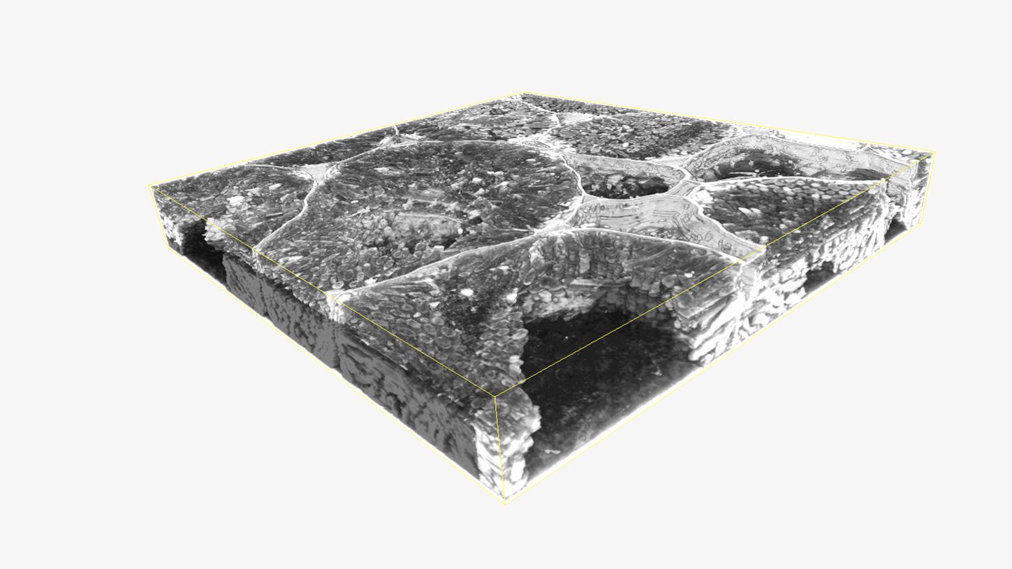
应用案例
具有原生质丝分布的根瘤连续切片的三维重构
![植物和细菌之间的共生关系 植物和细菌之间的共生关系]({"xsmall":"https://www.zeiss.com/content/dam/rms/reference-master/applications/life-sciences/volume-em/array-tomography/root-nodules-correlative-array-tomography.jpg/_jcr_content/renditions/original.image_file.100.100.file/root-nodules-correlative-array-tomography.jpg","small":"https://www.zeiss.com/content/dam/rms/reference-master/applications/life-sciences/volume-em/array-tomography/root-nodules-correlative-array-tomography.jpg/_jcr_content/renditions/original.image_file.360.360.file/root-nodules-correlative-array-tomography.jpg","medium":"https://www.zeiss.com/content/dam/rms/reference-master/applications/life-sciences/volume-em/array-tomography/root-nodules-correlative-array-tomography.jpg/_jcr_content/renditions/original.image_file.768.768.file/root-nodules-correlative-array-tomography.jpg","large":"https://www.zeiss.com/content/dam/rms/reference-master/applications/life-sciences/volume-em/array-tomography/root-nodules-correlative-array-tomography.jpg/_jcr_content/renditions/original./root-nodules-correlative-array-tomography.jpg","xlarge":"https://www.zeiss.com/content/dam/rms/reference-master/applications/life-sciences/volume-em/array-tomography/root-nodules-correlative-array-tomography.jpg/_jcr_content/renditions/original./root-nodules-correlative-array-tomography.jpg","xxlarge":"https://www.zeiss.com/content/dam/rms/reference-master/applications/life-sciences/volume-em/array-tomography/root-nodules-correlative-array-tomography.jpg/_jcr_content/renditions/original./root-nodules-correlative-array-tomography.jpg","max":"https://www.zeiss.com/content/dam/rms/reference-master/applications/life-sciences/volume-em/array-tomography/root-nodules-correlative-array-tomography.jpg/_jcr_content/renditions/original./root-nodules-correlative-array-tomography.jpg"})
联系我们
![植物和细菌之间的共生关系 由美国特拉华大学的D. Sherrier、J. Caplan和S. Modla提供。 植物和细菌之间的共生关系 由美国特拉华大学的D. Sherrier、J. Caplan和S. Modla提供。]({"xsmall":"https://www.zeiss.com/content/dam/rms/reference-master/applications/life-sciences/volume-em/array-tomography/root-nodules-correlative-array-tomography.jpg/_jcr_content/renditions/original.image_file.100.100.file/root-nodules-correlative-array-tomography.jpg","small":"https://www.zeiss.com/content/dam/rms/reference-master/applications/life-sciences/volume-em/array-tomography/root-nodules-correlative-array-tomography.jpg/_jcr_content/renditions/original.image_file.360.360.file/root-nodules-correlative-array-tomography.jpg","medium":"https://www.zeiss.com/content/dam/rms/reference-master/applications/life-sciences/volume-em/array-tomography/root-nodules-correlative-array-tomography.jpg/_jcr_content/renditions/original.image_file.768.768.file/root-nodules-correlative-array-tomography.jpg","large":"https://www.zeiss.com/content/dam/rms/reference-master/applications/life-sciences/volume-em/array-tomography/root-nodules-correlative-array-tomography.jpg/_jcr_content/renditions/original./root-nodules-correlative-array-tomography.jpg","xlarge":"https://www.zeiss.com/content/dam/rms/reference-master/applications/life-sciences/volume-em/array-tomography/root-nodules-correlative-array-tomography.jpg/_jcr_content/renditions/original./root-nodules-correlative-array-tomography.jpg","xxlarge":"https://www.zeiss.com/content/dam/rms/reference-master/applications/life-sciences/volume-em/array-tomography/root-nodules-correlative-array-tomography.jpg/_jcr_content/renditions/original./root-nodules-correlative-array-tomography.jpg","max":"https://www.zeiss.com/content/dam/rms/reference-master/applications/life-sciences/volume-em/array-tomography/root-nodules-correlative-array-tomography.jpg/_jcr_content/renditions/original./root-nodules-correlative-array-tomography.jpg"})
由美国特拉华大学的D. Sherrier、J. Caplan和S. Modla提供。
植物和细菌之间的共生关系
洞察根瘤中的细菌对植物健康和状态的影响
植物的根系网是植物获取全部水分和养分的途径,而水分和养分是所有植物生长的关键成分。为了促进植物健康成长并提升产量,探索整个根系网并了解外部微生物的影响至关重要。为了研究植物与根瘤中细菌之间的共生关系,必须了解根瘤和细菌的分布,而荧光和高分辨率结构组合评估对于详细了解这一点尤为重要。
Correlative Array Tomography能够叠加荧光和结构数据,从而实现根瘤和细菌分布的可视化。